
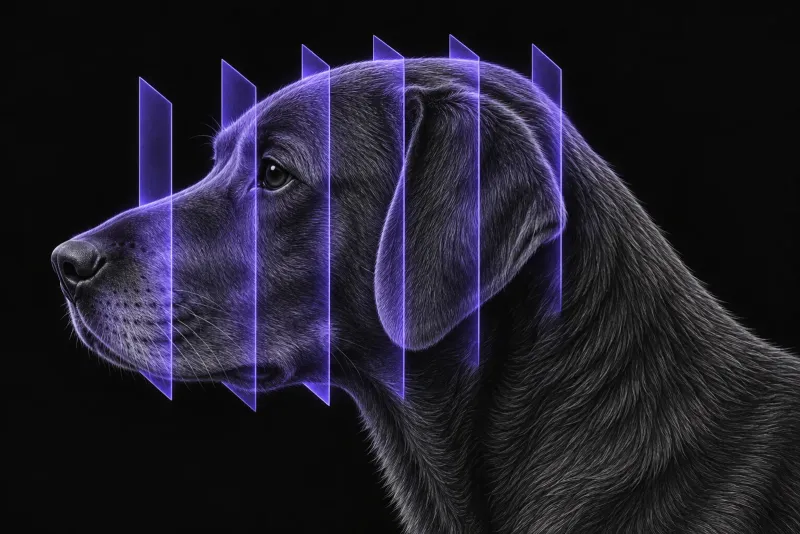
Why head trauma deserves a structured work-up
Traumatic skull fractures are relatively frequent in dogs. In a multicentre study of 91 dogs with skull fractures confirmed on CT (Amengual-Batle et al., Journal of Veterinary Internal Medicine, 2020), the most common known causes were road traffic accidents (26%) and dog attacks (21%). The cause was unknown in about 17% of dogs. Chihuahuas and Yorkshire Terriers were the two most commonly affected pedigree breeds.
The neurological examination comes first. In 72 dogs with head trauma (Sharma and Holowaychuk, Journal of Veterinary Emergency and Critical Care, 2015), a decreasing modified Glasgow Coma Scale (MGCS) score was the strongest predictor of non-survival. A score of 11 or less was 84% sensitive and 73% specific for non-survival. CT adds structural information to that assessment. A CT-based prognostic score (Rapoport et al., The Veterinary Journal, 2020) has been described as a complementary tool for serial clinical and neurological evaluation.
When CT is indicated
CT is described as the imaging modality of choice for acute head trauma (Hecht et al., Frontiers in Veterinary Science, 2021), and it gives excellent bony resolution. In practice, CT is usually worth discussing when a dog has:
- neurological deficits or a falling MGCS score
- a palpable skull deformity
- facial fractures that may need surgical repair
Include the cervical spine. At least 5% of dogs and cats with skull fractures also had cervical vertebral fractures or luxations. The authors stress imaging this region, because lesions at several sites can be hard to identify clinically.
What CT shows
In 27 dogs admitted within 72 hours of head trauma that had head CT (Chai et al., American Journal of Veterinary Research, 2017), the findings were:
| Finding | Dogs |
|---|---|
| Cranial vault fracture, parenchymal abnormality, or both | 24 of 27 (89%) |
| Intracranial haemorrhage | 16 (59%) |
| Cranial vault fracture | 15 (56%) |
| Midline shift | 14 (52%) |
| Lateral ventricle asymmetry | 12 (44%) |
| Hydrocephalus | 7 (26%) |
| Facial bone fractures only | 3 (11%) |
In the 91 dogs of the multicentre study, the cranial vault was fractured in 72 (79.1%), the face in 45 (49.5%) and the base of the cranium in 18 (19.8%). Neurological signs were present in 57 dogs (62.6%).
CT and prognosis: useful, but the evidence is mixed
- The Koret CT score. From the 27-dog study, a 7-point score was built: 1 point each for haemorrhage, midline shift or lateral ventricle asymmetry, cranial vault fracture and depressed fracture, and 3 points for an infratentorial lesion. In a later study of 35 dogs, a higher score was associated with lower short- and long-term survival. A score of 3 or less was associated with survival with 85% sensitivity and 100% specificity.
- A negative study. In 40 dogs scanned within 72 hours (Wyatt et al., Veterinary Radiology & Ultrasound) and graded on a modified imaging scale, no CT feature was associated with survival to discharge or with immediate post-traumatic seizures. Thirty dogs (75%) survived to discharge.
Where CT stops: MRI
In 50 dogs with traumatic brain injury that had MRI within 14 days (Beltran et al., Journal of Veterinary Internal Medicine, 2014), MRI grade correlated negatively with the MGCS score. Midline shift and the percentage of intraparenchymal lesions correlated with worse follow-up scores, and 10% of dogs developed post-traumatic epilepsy.
MRI can also show fractures. In a cadaver model, MRI agreed with CT on the presence or absence of a fracture in 93.5% of assessments overall, but in 87.6% of the post-trauma studies.
Skull CT protocol at a glance
Head CT for trauma relies on CT’s bony detail, and the scan should take in the cervical spine as well as the skull. Facial fractures travel in groups, so the authors of a CT study of 165 dogs (De Paolo et al., Frontiers in Veterinary Science, 2020) recommend a thorough CT evaluation of the whole region, even when the visible fractures look superficial. When MRI is used for head trauma, adding a proton-density-weighted sequence is recommended.
What the radiologist looks at
- Where the fracture is. Location matters more than the presence of a fracture. Across dogs and cats, a cranial vault fracture was a strong risk factor for neurological signs, especially when depressed. Animals with fractures of the face only were less likely to have neurological signs and had no secondary intracranial changes.
- Fracture groups. In the 165-dog study, maxillary fractures came with many concurrent fractures of the midface, skull base and cranial vault. A fractured zygomatic bone tended to occur with fractures of the other orbital bones, and frontal or nasal bone fractures were often accompanied by skull base fractures. Vehicular trauma produced more severely displaced and comminuted fractures than other causes.
- Each fracture described. Every fracture is described by region, fragmentation and displacement.
- The brain. Haemorrhage, midline shift, ventricular asymmetry, herniation and any infratentorial lesion, the findings that make up the Koret CT score.
What your report will include
Every Veheri case is read by a radiologist or neurologist, and when you order you can choose Diplomate-only reporting or the full specialist panel. Neurology reporting covers CT and MRI. You can chat directly with the specialist assigned to your case, and a simplified pet owner report helps you explain the findings to the owner.
When you take the report forward:
- Read the CT with the MGCS score. CT findings support a prognosis, but one study found no CT feature associated with survival, so serial neurological assessment still leads.
- A brain parenchyma question, rather than a bone question, makes MRI worth discussing once the patient is stable. In the 50-dog MRI study, MRI grade correlated negatively with the MGCS score.
- Talking to the owner: fractures alone are not a death sentence. The one-week case fatality rate in dogs with skull fractures was 16.5%. Death was more likely with cranial vault fractures, particularly fragmented or displaced ones, but most animals survived the first week.
What to include when you request a skull CT report
- signalment: age, breed and sex
- time since the trauma and the mechanism, such as road traffic accident, bite or fall
- MGCS score at admission and any change since
- neurological deficits, seizures and neck pain
- facial swelling, malocclusion or inability to open or close the mouth
- whether the cervical spine should be included
How Veheri can help
Veheri provides veterinary teleradiology and teleneurology for X-ray, CT and MRI, with expert support for CT scanning protocols, positioning and acquisition. Related reading: CT of the feline skull after trauma, CT of the canine pelvis after trauma and the role of emergency CT.
Sources
Each claim in this guide links to its source. The full citations:
- Amengual-Batle P, José-López R, Durand A, et al. Traumatic skull fractures in dogs and cats: a comparative analysis of neurological and computed tomographic features. J Vet Intern Med. 2020;34(5):1975-1985. doi:10.1111/jvim.15838
- Sharma D, Holowaychuk MK. Retrospective evaluation of prognostic indicators in dogs with head trauma: 72 cases (January-March 2011). J Vet Emerg Crit Care (San Antonio). 2015;25(5):631-9. doi:10.1111/vec.12328
- Rapoport K, Mateo I, Peery D, et al. The prognostic value of the Koret CT score in dogs following traumatic brain injury. Vet J. 2020;266:105563. doi:10.1016/j.tvjl.2020.105563
- Hecht S, Anderson KM, Castel A, et al. Agreement of magnetic resonance imaging with computed tomography in the assessment for acute skull fractures in a canine and feline cadaver model. Front Vet Sci. 2021;8:603775. doi:10.3389/fvets.2021.603775
- Chai O, Peery D, Bdolah-Abram T, et al. Computed tomographic findings in dogs with head trauma and development of a novel prognostic computed tomography-based scoring system. Am J Vet Res. 2017;78(9):1085-1090. doi:10.2460/ajvr.78.9.1085
- De Paolo MH, Arzi B, Pollard RE, et al. Craniomaxillofacial trauma in dogs. Part II: association between fracture location, morphology and etiology. Front Vet Sci. 2020;7:242. doi:10.3389/fvets.2020.00242
- Wyatt S, Llabres-Diaz F, Lee CY, Beltran E. Early CT in dogs following traumatic brain injury has limited value in predicting short-term prognosis. Vet Radiol Ultrasound. 62(2):181-189 (published online 2020). doi:10.1111/vru.12933
- Beltran E, Platt SR, McConnell JF, et al. Prognostic value of early magnetic resonance imaging in dogs after traumatic brain injury: 50 cases. J Vet Intern Med. 2014;28(4):1256-62. doi:10.1111/jvim.12368
Related reading
More imaging guides
All imaging guides
CT of the feline skull for head and maxillofacial trauma
Which skull, jaw and temporomandibular joint fractures CT finds in cats after head trauma, why radiographs miss them, and which findings change treatment.

CT of adrenal masses in dogs
What contrast CT shows in a dog with an adrenal mass, from caval invasion and adhesions to tumour type, and where its accuracy runs out before adrenalectomy.

Brain CT in dogs when MRI is not available
What brain CT can and cannot detect in dogs when MRI is not available, for masses, stroke, inflammation and head trauma, and when MRI or CSF is still needed.