
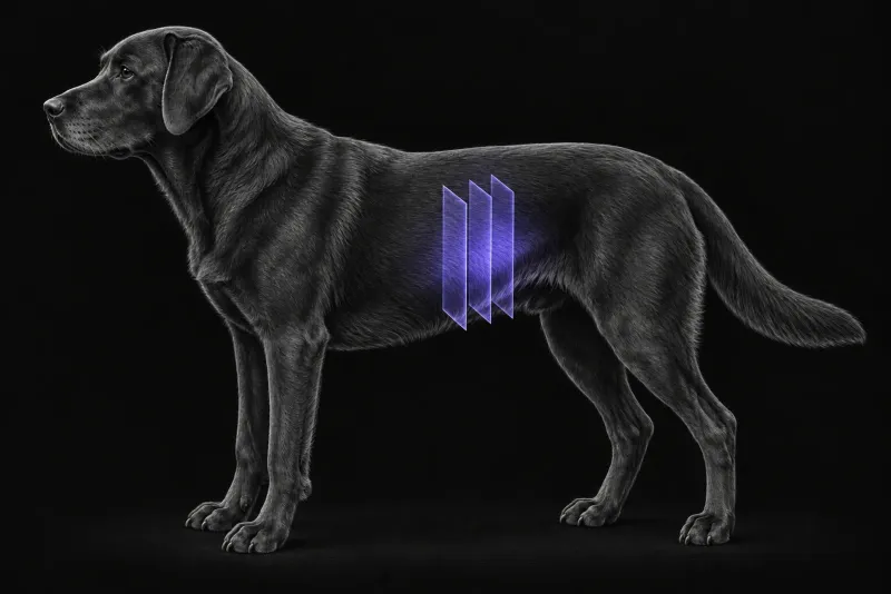
Why a splenic mass is a hard question
A splenic mass is roughly as likely to be benign as malignant. In 234 dogs with a splenic mass found on ultrasound or CT and a confirmed diagnosis (O’Byrne and Hosgood, Veterinary Record, 2019), 55% had malignant and 45% benign disease.
- Small-breed dogs were 2.3 times more likely than large-breed dogs to have benign disease.
- When small dogs did have malignant disease, they were one-third as likely as large-breed dogs to have haemangiosarcoma.
The owner’s decision often rests on the chance of malignancy, so it is natural to hope that CT can settle it. The evidence says CT can shift that probability but not replace histology.
What CT shows at the mass
| Study | Main finding |
|---|---|
| Dual-phase CT, 31 splenic masses, 18 malignant (Jones et al., Veterinary Radiology & Ultrasound, 2016) | Substantial overlap between malignant and non-malignant masses. Haemangiosarcoma and nodular hyperplasia most often showed marked enhancement in the early phase that persisted in the delayed phase. Most haematomas enhanced only slightly in the early phase. All haematomas and 77% of haemangiosarcomas showed contrast accumulation compatible with active haemorrhage. |
| Triple-phase CT, 42 dogs (Kutara et al., Journal of Veterinary Medical Science, 2017) | Nodular hyperplasia enhanced homogeneously and normally in all phases. Haemangiosarcoma showed either homogeneous poor enhancement in all phases, or heterogeneous marked enhancement in the arterial and portal venous phases. Haematoma and undifferentiated sarcoma showed heterogeneous normal enhancement. |
| 57 dogs with splenectomy (Lee et al., Journal of Veterinary Science, 2018) | Mean pre-contrast attenuation was lower in malignant (40.3 ± 5.9 HU) than in benign tumours (52.8 ± 6.8 HU). |
| Focal splenic lesions, decision tree (Burti et al., Frontiers in Veterinary Science, 2022) | Sarcomas were large, cystic and enhanced little. Nodular hyperplasia and other benign lesions were small, solid and enhanced strongly. Round cell tumours had no distinctive features. Accuracy was 0.89 for sarcoma and 0.79 for benign lesions; round cell tumours could not be classified. |
The studies are small and used different contrast phases, and they do not fully agree. The dual-phase study found no other significant quantitative differences between malignant and non-malignant masses. It concluded that dual-phase CT gives limited specific information. The authors of the decision-tree study state that a definitive diagnosis needs microscopic examination of the spleen.
Nodules that look worrying but often are not
Extramedullary haematopoiesis is common in the canine spleen. In 89 dogs with a cytological diagnosis of it and three-phase CT (Cordella et al., Veterinary Radiology & Ultrasound, 2020), the spleen showed:
- multiple nodules in 62%
- a mass in 16%
- diffuse heterogeneous parenchyma in 13%
- a normal appearance in 9%
Most lesions were hyperattenuating to the spleen in the arterial (64%) and portal (66%) phases, but only 45% in the interstitial phase. A hyperattenuating nodule is therefore not on its own a sign of malignancy.
Where CT adds most: staging
If haemangiosarcoma is possible, the wider scan is often more useful than the spleen itself.
- Lungs. In 536 dogs with non-pulmonary malignancy (Lamb et al., Veterinary Radiology & Ultrasound, 2019), multiple pulmonary nodules were present at first CT in 24 of 58 dogs with haemangiosarcoma (41%), the highest proportion of any tumour type studied. A solitary nodule was an indeterminate finding.
- Skeletal muscle. In 61 dogs with haemangiosarcoma that had whole-body CT (Carloni et al., Journal of Veterinary Internal Medicine, 2019), skeletal muscle metastases were found in 15 (24.6%), and all of them also had metastases elsewhere. Nine of those 15 dogs were lame or reluctant to move, against none of the 42 dogs without muscle metastases. The authors recommend whole-body CT for staging.
- Liver and abdomen on ultrasound. In 99 dogs with haemoperitoneum from a ruptured splenic tumour (Ramirez et al., Journal of the American Veterinary Medical Association, 2024), ultrasound liver lesions were not associated with metastasis on liver biopsy (sensitivity 19%, specificity 82%). They were associated with a higher likelihood of a benign splenic tumour. Ultrasound missed 82% of intra-abdominal lesions beyond the liver and spleen. The authors suggest whole-body CT or MRI may be more warranted for staging.
Spleen CT protocol at a glance
The studies above used contrast CT in two or three phases, from dual-phase imaging to triple-phase imaging. Pre-contrast images matter too, because pre-contrast attenuation differed between malignant and benign tumours. When haemangiosarcoma is possible, whole-body CT is recommended for staging.
What the radiologist looks at
- Size, structure and enhancement. Large, cystic, poorly enhancing masses lean towards sarcoma, and small, solid, strongly enhancing lesions towards benign disease, in the decision-tree study.
- Pre-contrast attenuation. Lower values were seen in malignant tumours.
- Signs of active haemorrhage. These are not specific: they were seen in both haematomas and haemangiosarcomas.
- Hyperattenuating nodules. These may be extramedullary haematopoiesis rather than metastases.
- The rest of the body. Lungs, skeletal muscle and abdominal organs, because multiple pulmonary nodules and muscle metastases change staging.
What your report will include
Every Veheri case is read by a radiologist or neurologist, and when you order you can choose Diplomate-only reporting or the full specialist panel. You can chat directly with the specialist assigned to your case, and a simplified pet owner report helps you explain the findings to the owner.
When you take the report forward:
- Histology is still needed. Benign and malignant masses overlap on CT, and a definitive diagnosis needs microscopic examination.
- Reported patterns support a probability, not a diagnosis. They come from small cohorts with different protocols.
- A solitary pulmonary nodule is an indeterminate finding, unlike multiple nodules.
- Liver lesions on ultrasound in a dog with haemoperitoneum were not associated with metastasis on biopsy.
- Talking to the owner: a splenic mass is roughly as likely to be benign as malignant, and CT shifts that probability rather than settling it.
What to include when you request a splenic CT report
- signalment: age, breed and sex
- body size, since small-breed dogs are more likely to have benign disease
- whether there is haemoperitoneum, and the patient’s stability and transfusion history
- ultrasound findings in the spleen and liver
- lameness or reluctance to move, which may point to muscle metastases
- any other known tumour or previous splenic sampling
- whether thorax and whole-body imaging should be included in the same study
How Veheri can help
Veheri’s radiologists report veterinary CT studies remotely for clinics, and can support your team with CT scanning protocols for abdominal and staging CT. For related reading, see multiphase CT of liver masses in dogs and the role of emergency CT in veterinary practice.
Sources
Each claim in this guide links to its source. The full citations:
- O'Byrne K, Hosgood G. Splenic mass diagnosis in dogs undergoing splenectomy according to breed size. Vet Rec. 2019;184(20):620. doi:10.1136/vr.104983
- Jones ID, Lamb CR, Drees R, et al. Associations between dual-phase computed tomography features and histopathologic diagnoses in 52 dogs with hepatic or splenic masses. Vet Radiol Ultrasound. 2016;57(2):144-53. doi:10.1111/vru.12336
- Kutara K, Seki M, Ishigaki K, et al. Triple-phase helical computed tomography in dogs with solid splenic masses. J Vet Med Sci. 2017;79(11):1870-1877. doi:10.1292/jvms.17-0253
- Lee M, Park J, Choi H, et al. Presurgical assessment of splenic tumors in dogs: a retrospective study of 57 cases (2012-2017). J Vet Sci. 2018;19(6):827-834. doi:10.4142/jvs.2018.19.6.827
- Burti S, Zotti A, Bonsembiante F, et al. A machine learning-based approach for classification of focal splenic lesions based on their CT features. Front Vet Sci. 2022;9:872618. doi:10.3389/fvets.2022.872618
- Cordella A, Caldin M, Bertolini G. Splenic extramedullary hematopoiesis in dogs is frequently detected on multiphase multidetector-row CT as hypervascular nodules. Vet Radiol Ultrasound. 2020;61(5):512-518. doi:10.1111/vru.12872
- Lamb CR, Whitlock J, Foster-Yeow ATL. Prevalence of pulmonary nodules in dogs with malignant neoplasia as determined by CT. Vet Radiol Ultrasound. 2019;60(3):300-305. doi:10.1111/vru.12723
- Carloni A, Terragni R, Morselli-Labate AM, et al. Prevalence, distribution, and clinical characteristics of hemangiosarcoma-associated skeletal muscle metastases in 61 dogs: a whole body computed tomographic study. J Vet Intern Med. 2019;33(2):812-819. doi:10.1111/jvim.15456
- Ramirez AR, Stewart SD, Khanna C. Ultrasonographic liver nodules are more often benign lesions in dogs with hemoperitoneum secondary to splenic tumor rupture. J Am Vet Med Assoc. 2024;262(11):1499-1503. doi:10.2460/javma.24.04.0254
Related reading
More imaging guides
All imaging guides
CT of adrenal masses in dogs
What contrast CT shows in a dog with an adrenal mass, from caval invasion and adhesions to tumour type, and where its accuracy runs out before adrenalectomy.

Brain CT in dogs when MRI is not available
What brain CT can and cannot detect in dogs when MRI is not available, for masses, stroke, inflammation and head trauma, and when MRI or CSF is still needed.
CT of the canine elbow for medial coronoid disease
When CT is worth it for medial coronoid disease in dogs, what it finds that radiographs miss, and where arthroscopy still gives the better answer.