
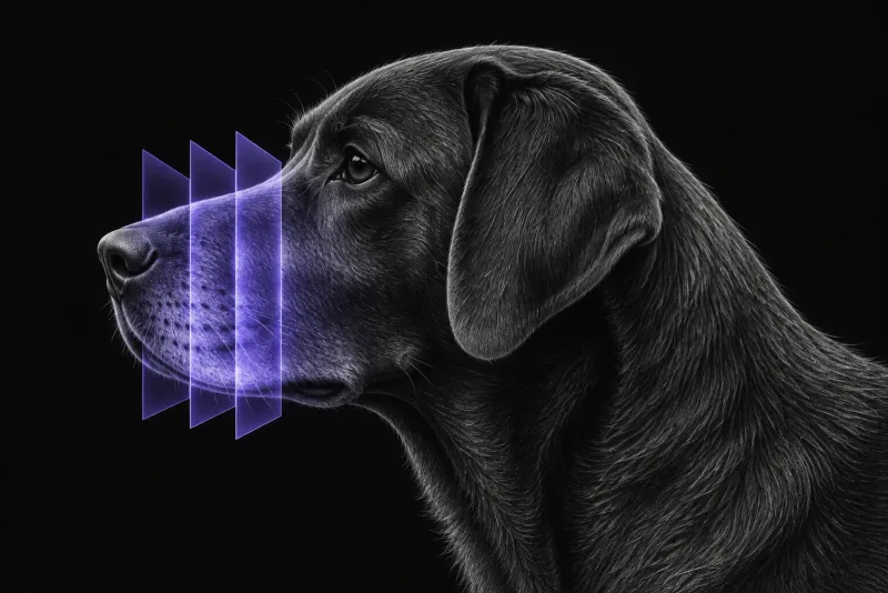
Why oral tumours are staged with CT
Oral tumours in dogs often involve bone. In a 15-year review of 381 dogs with histologically confirmed oral tumours (Goldschmidt et al., Journal of the American Veterinary Medical Association, 2023), 348 had CT of the skull and neck, and bone lysis was present in 74.4% of tumours. Squamous cell carcinoma, sarcomas and tumours larger than 2 cm (T2 to T3) had significantly more lysis than odontogenic tumours and tumours smaller than 2 cm (T1).
Radiographs underestimate this. In 21 dogs with maxillary masses (Ghirelli et al., Journal of Veterinary Dentistry, 2013), bony changes were seen in 80.9% of cases on radiographs and 95.2% on CT. Invasion of neighbouring structures was seen in 30% of cases on radiographs but in 90.4% on CT. These structures included:
- the nasal cavity
- the frontal and sphenoidal sinuses
- the orbit and the maxillary recess
- the nasopharynx
The lymph nodes: where CT is weakest
Cervical lymph node metastasis alters treatment and prognosis (Goldschmidt et al., PLOS ONE, 2023), but CT appearance alone is not a reliable test.
| Study | Finding |
|---|---|
| 40 dogs with oral or nasal cancer, all mandibular and medial retropharyngeal nodes removed (Skinner et al., Veterinary and Comparative Oncology, 2018) | Metastasis in 16 dogs. CT sensitivity was 12.5% for mandibular and 10.5% for medial retropharyngeal nodes; specificity was 91.1% and 96.7%. No single CT finding predicted metastasis. |
| 39 dogs, indirect CT lymphangiography, then bilateral node removal | A sentinel node was found in 38 (97%). In the 13 dogs with metastasis, CT lymphangiography identified the draining lymphocentre in all. Lymphangiography features alone could not predict metastasis. |
| 74 dogs (77 tumours), CT lymphangiography (Chrostek et al., Veterinary and Comparative Oncology, 2026) | Contralateral or bilateral drainage occurred in over 55% of rostral tumours. Of 10 dogs with node metastasis, 4 (40%) had metastasis beyond the ipsilateral lateral mandibular node, and 3 had it only in the medial retropharyngeal nodes. |
The two contrast-enhanced CT studies disagree. One found good accuracy for contrast-enhanced CT features; the other found very low sensitivity and concluded that CT cannot be relied on alone.
In the 381-dog series, node metastasis was diagnosed by CT or cytology in 10.7% of non-odontogenic tumours and 0% of odontogenic tumours. Melanoma, squamous cell carcinoma and T3 tumours had the highest prevalence. Bone lysis was not associated with cervical metastasis. Cytology was not performed in most cases and often included only a single mandibular node, so the authors believe this underestimates metastasis.
Distant staging
In the same population, screened for distant disease (Goldschmidt et al., Journal of the American Veterinary Medical Association, 2023), thoracic metastasis was found in 4.9% of oral tumours (0.9% of odontogenic and 6.5% of non-odontogenic). Melanoma and multilobular osteochondrosarcoma were most at risk. Metastasis and major findings were diagnosed more often with thoracic CT than with radiographs. In the 115 dogs that had head CT, node cytology, thoracic screening and abdominal ultrasound, at least one test found metastasis or a major finding of prognostic significance in 27.8%.
Oral CT protocol at a glance
Staging CT for an oral tumour covers the skull and neck, as in the 381-dog series. Adding the thorax to the same anaesthetic is usually worth discussing, because thoracic CT found metastasis more often than radiographs. Indirect CT lymphangiography can map the draining lymphocentre, which varies from dog to dog.
What the radiologist looks at
- Signs that point to malignancy. In 28 dogs with 31 oral masses (Lee et al., Veterinary Radiology & Ultrasound, 2021), these were large size, heterogeneous contrast enhancement, bone lysis, tooth loss and ipsilateral mandibular lymphadenopathy.
- Benign tumours can look aggressive. In the same study, bone lysis also occurred in acanthomatous ameloblastoma, giant cell granuloma and plasmacytoma. The authors found considerable overlap between tumour types and between benign and malignant tumours.
- Odontogenic tumours. In 29 dogs (Amory et al., Veterinary Radiology & Ultrasound, 2014), the most common CT features were an association with multiple teeth (96.4%), contrast enhancement (96.3%), alveolar bone lysis (93.1%) and tooth displacement (85.2%). CT showed extension beyond the oral cavity more often than dental radiographs. One of four fibromatous epulides was not visible on CT.
- Both sides of the neck. Node size and enhancement do not reliably detect metastasis, and drainage can be contralateral or bilateral.
- Incidental findings. Head CT found major incidental findings in 4.6% of scans, mainly other tumours outside the mouth. The risk rose with age.
What your report will include
Every Veheri case is read by a radiologist or neurologist, and when you order you can choose Diplomate-only reporting or the full specialist panel. You can chat directly with the specialist assigned to your case, and a simplified pet owner report helps you explain the findings to the owner.
When you take the report forward:
- Histology is still needed. CT features overlap between benign and malignant tumours, and the authors concluded that histology remains necessary for a definitive diagnosis.
- Sample the nodes. CT lymphangiography maps the nodes to sample but does not replace cytology or histology. Its authors recommend cytological or histological sampling of the sentinel node before treatment. The 381-dog series authors highly recommend guided node sampling, especially for melanoma, squamous cell carcinoma and T2 to T3 tumours.
- Fine dental detail. Dental radiographs show tooth destruction more often than CT, so the two answer different questions.
What to include when you request an oral CT report
- signalment: age, breed and sex
- location and size of the mass, and whether it is lateral or near the midline
- biopsy result, if already available
- results of any node palpation or cytology, and which node was sampled
- whether the thorax should be included
- the treatment being considered, such as surgery or radiotherapy
- any previous oral surgery or extractions at the site
How Veheri can help
Veheri provides veterinary teleradiology for X-ray, CT and MRI, with expert support for CT scanning protocols, positioning and acquisition. Related reading: CT of the feline oral cavity, thoracic CT for pulmonary metastases in dogs and oncology imaging for staging and planning.
Sources
Each claim in this guide links to its source. The full citations:
- Goldschmidt S, Soltero-Rivera M, Quiroz A, et al. The diagnostic yield of preoperative screening for oral cancer in dogs over 15 years, part 1: locoregional screening. J Am Vet Med Assoc. 2023;261(S2):S14-S23. doi:10.2460/javma.23.05.0299
- Ghirelli CO, Villamizar LA, Pinto AC. Comparison of standard radiography and computed tomography in 21 dogs with maxillary masses. J Vet Dent. 2013;30(2):72-6. doi:10.1177/089875641303000201
- Lee S, Jang Y, Lee G, et al. CT features of malignant and benign oral tumors in 28 dogs. Vet Radiol Ultrasound. 2021;62(5):549-556. doi:10.1111/vru.12996
- Amory JT, Reetz JA, Sanchez MD, et al. Computed tomographic characteristics of odontogenic neoplasms in dogs. Vet Radiol Ultrasound. 2014;55(2):147-58. doi:10.1111/vru.12101
- Goldschmidt S, Stewart N, Ober C, et al. Contrast-enhanced and indirect computed tomography lymphangiography accurately identifies the cervical lymphocenter at risk for metastasis in pet dogs with spontaneously occurring oral neoplasia. PLoS One. 2023;18(3):e0282500. doi:10.1371/journal.pone.0282500
- Skinner OT, Boston SE, Giglio RF, et al. Diagnostic accuracy of contrast-enhanced computed tomography for assessment of mandibular and medial retropharyngeal lymph node metastasis in dogs with oral and nasal cancer. Vet Comp Oncol. 2018;16(4):562-570. doi:10.1111/vco.12415
- Chrostek E, Lawrence J, Arzi B, et al. Sentinel lymph node mapping identifies variable cervical drainage and metastatic nodes outside traditional sampling sites in canine oromaxillofacial tumours. Vet Comp Oncol. 2026 (online ahead of print). doi:10.1111/vco.70090
- Goldschmidt S, Soltero-Rivera M, Quiroz A, et al. The diagnostic yield of preoperative screening for oral cancer in dogs over 15 years, part 2: distant screening. J Am Vet Med Assoc. 2023;261(S2):S24-S33. doi:10.2460/javma.23.05.0300
Related reading
More imaging guides
All imaging guides
CT of the feline oral cavity for squamous cell carcinoma staging
How CT stages oral squamous cell carcinoma in cats, what bone lysis and orbital extension look like, how often nodes are involved, and where CT falls short.

CT of adrenal masses in dogs
What contrast CT shows in a dog with an adrenal mass, from caval invasion and adhesions to tumour type, and where its accuracy runs out before adrenalectomy.

Brain CT in dogs when MRI is not available
What brain CT can and cannot detect in dogs when MRI is not available, for masses, stroke, inflammation and head trauma, and when MRI or CSF is still needed.