
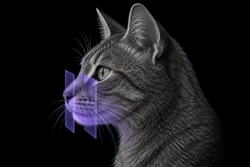
Why oral tumours in cats need cross-sectional staging
Squamous cell carcinoma (SCC) is the most common oral malignancy in cats, and it is highly locally invasive. In 20 cats treated by total glosso-mandibulectomy (Iwata et al., Veterinary and Comparative Oncology, 2025), the histopathological margin was the only factor associated with progression-free and overall survival on univariate analysis. Knowing how far the tumour extends before surgery or radiotherapy is therefore central to planning.
Not every oral mass is SCC. In 60 cats treated by maxillectomy for an oral tumour (Liptak et al., Veterinary and Comparative Oncology, 2021), 19 (31.7%) had benign tumours and 41 (68.3%) malignant ones. Imaging stages the mass, but histology names it.
What CT shows in feline oral SCC
In 18 cats with oral SCC (Gendler et al., Journal of the American Veterinary Medical Association, 2010), CT located the tumour in:
- the sublingual or lingual region (7 cats)
- the maxilla (5)
- the buccal mucosa (4) and mandible (4)
- the pharyngeal mucosa (2), soft palate (1) and lip (1)
Key findings:
| CT finding | What the evidence shows |
|---|---|
| Osteolysis | Osteolytic mass lesions in 9 of 18 cats (Gendler et al.) |
| Orbital extension | The most common extension of maxillary masses (5 cats) (Gendler et al.) |
| Contrast enhancement | Heterogeneous enhancement was the most common pattern (8 of 18) (Gendler et al.) |
| Periosteal reaction | Maxillary SCC showed severe cortical destruction with irregular, “pumice stone-like” periosteal reaction (4 maxillary masses in a series of 10 cats) (Strohmayer et al., Frontiers in Veterinary Science, 2020) |
| Deep bone invasion | Marked, extensive and deep bone invasion on histology in most horses and cats in the same study (Strohmayer et al.) |
CT and oral examination agreed on tumour location at every site except the soft palate, where CT identified 1 cat and oral examination 4. A careful oral examination under anaesthesia remains part of staging.
None of the quantitative CT features, including mass size, attenuation and lymph node width, was correlated with survival time. CT phenotypes were also unrelated to histological grade. CT describes extent; it does not grade or predict.
Lymph nodes and lungs
Regional spread is not rare. In 49 cats with advanced oral SCC assessed by mandibular lymph node cytology and three-view thoracic radiographs (Soltero-Rivera et al., Journal of Feline Medicine and Surgery, 2014):
- mandibular lymph node metastasis was present in 31% (15 of 49)
- possible thoracic metastasis was seen in 10% (5 of 49)
- of the nodal metastases, 8 came from maxillary, 4 from mandibular, 2 from sublingual and 1 from caudal pharyngeal tumours
The authors note that both rates were higher than most earlier reports. CT can show lymph node enlargement, but node size alone is not proof of metastasis. In a PET/CT study of 12 cats (Randall et al., Veterinary and Comparative Oncology, 2016), two of three cytologically confirmed metastases were equivocal on CT.
Where CT underestimates the tumour
CT is less reliable for soft-tissue infiltration than for bone:
- Soft-tissue infiltrative tumours that were subtle and ill defined on CT were highly visible and more extensive on FDG-PET/CT. Tumours invading bone were more similar in extent on both methods.
- In lingual and laryngeal SCC, most cats had a large region of high FDG uptake outside the CT tumour volume (Yoshikawa et al., Veterinary Radiology & Ultrasound, 2013).
Oral cavity CT protocol at a glance
Staging uses contrast-enhanced CT of the head, with the regional lymph nodes in the field, as in the CT study of 18 cats. When CT is performed under anaesthesia for the oral mass, including the thorax is worth discussing, so the head, neck and lungs are staged in one session. The same anaesthetic is a chance for a careful oral examination.
What the radiologist looks at
- Bone. Osteolysis, cortical destruction and periosteal reaction, which were common in maxillary SCC.
- Extension. Into the orbit, the most common extension of maxillary masses, and into neighbouring structures.
- Soft-tissue margins. Read with caution in tongue and sublingual tumours, where CT can underestimate the tumour volume.
- Lymph nodes. Size and appearance of the regional nodes, knowing that metastases can look equivocal on CT.
- The lungs. When the thorax is included, for possible metastasis.
What your report will include
Every Veheri case is read by a radiologist or neurologist, and when you order you can choose Diplomate-only reporting or the full specialist panel. You can chat directly with the specialist assigned to your case, and a simplified pet owner report helps you explain the findings to the owner.
When you take the report forward:
- Sample the mass. 19 of 60 cats treated by maxillectomy (31.7%) had benign tumours, so histology is still needed.
- Sample the nodes. Cytology of the regional nodes is usually still worth planning, whatever their size on CT, because nodal metastasis was present in 31%.
- Soft-tissue extent may be larger than on CT. For tongue and sublingual tumours in particular, the CT margin may not show the full extent of disease. This matters for surgical margins and radiotherapy fields.
- Talking to the owner: the histopathological margin was the only factor associated with survival after glosso-mandibulectomy, and CT features did not predict survival time.
What to include when you request an oral CT report
- signalment: age, breed and sex
- location of the mass on oral examination, including the soft palate
- duration, dysphagia, ptyalism, facial swelling or exophthalmos
- biopsy or cytology results, if available
- mandibular lymph node findings on palpation or cytology
- the planned treatment, so the report focuses on surgical margins, orbital and nasal extension or radiotherapy volumes
How Veheri can help
Veheri’s radiologists report veterinary CT studies remotely for clinics, and can support your team with CT scanning protocols for head and neck staging. For related reading, see CT of the canine oral cavity and oncology imaging for staging and planning.
Sources
Each claim in this guide links to its source. The full citations:
- Iwata T, Ichimata M, Fujita A, et al. The outcomes and surgical complications of cats with oral squamous cell carcinoma treated with total glosso-mandibulectomy: 20 cases (2008-2022). Vet Comp Oncol. 2025;23(2):178-186. doi:10.1111/vco.13043
- Liptak JM, Thatcher GP, Mestrinho LA, et al. Outcomes of cats treated with maxillectomy: 60 cases. A Veterinary Society of Surgical Oncology retrospective study. Vet Comp Oncol. 2021;19(4):641-650. doi:10.1111/vco.12634
- Gendler A, Lewis JR, Reetz JA, Schwarz T. Computed tomographic features of oral squamous cell carcinoma in cats: 18 cases (2002-2008). J Am Vet Med Assoc. 2010;236(3):319-25. doi:10.2460/javma.236.3.319
- Strohmayer C, Klang A, Kneissl S. Computed tomographic and histopathological characteristics of 13 equine and 10 feline oral and sinonasal squamous cell carcinomas. Front Vet Sci. 2020;7:591437. doi:10.3389/fvets.2020.591437
- Soltero-Rivera MM, Krick EL, Reiter AM, et al. Prevalence of regional and distant metastasis in cats with advanced oral squamous cell carcinoma: 49 cases (2005-2011). J Feline Med Surg. 2014;16(2):164-9. doi:10.1177/1098612X13502975
- Randall EK, Kraft SL, Yoshikawa H, LaRue SM. Evaluation of 18F-FDG PET/CT as a diagnostic imaging and staging tool for feline oral squamous cell carcinoma. Vet Comp Oncol. 2016;14(1):28-38. doi:10.1111/vco.12047
- Yoshikawa H, Randall EK, Kraft SL, Larue SM. Comparison between 2-(18)F-fluoro-2-deoxy-D-glucose positron emission tomography and contrast-enhanced computed tomography for measuring gross tumor volume in cats with oral squamous cell carcinoma. Vet Radiol Ultrasound. 2013;54(3):307-13. doi:10.1111/vru.12016
Related reading
More imaging guides
All imaging guides
CT staging of oral tumours in dogs
How CT stages canine oral tumours, from bone invasion to the mandibular and medial retropharyngeal nodes, and why node sampling is still needed.
CT of the feline interscapular region for injection-site sarcoma
What contrast CT shows in feline injection-site sarcoma, how it changes surgical and radiotherapy planning, and where imaging cannot replace histology.
Thoracic CT in cats for lung tumours, airway and interstitial disease
What thoracic CT adds over radiographs in cats with lung tumours, bronchial or interstitial disease, how anaesthesia affects the lungs, and where CT stops.