
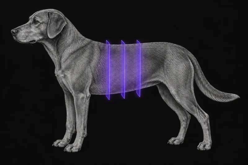
The diagnosis is biochemical; CT is for localisation and staging
Insulinoma is the most common pancreatic tumour in dogs. According to a 2022 review of canine insulinoma (Buishand, Veterinary Sciences), more than 95% are considered malignant because they almost always metastasise.
The diagnosis rests on blood tests, not imaging: hypoglycaemia together with an insulin concentration within or above the upper end of the reference interval. A blood glucose below 3.5 mmol/L with a plasma insulin above 10 μU/mL at the same time is characteristic.
Imaging then answers the surgical questions. In the same review:
- most primary tumours are nodules under 2.5 cm, usually in the left or right pancreatic limb rather than the body
- multiple primary tumours occur in 4 to 14% of cases
- metastases, mainly in the abdominal lymph nodes and liver, are already visible at surgery in 40 to 50% of dogs
Knowing the location before surgery helps choose between open and laparoscopic surgery, and between enucleation and partial pancreatectomy.
Why CT is the preferred staging method
The 2022 review cites a sensitivity of 36% for transabdominal ultrasound in detecting canine insulinoma, and notes that ultrasound alone is insufficient for staging. It recommends contrast-enhanced CT as the preferred staging method. In three dogs scanned with dual-phase CT angiography (Mai and Cáceres, Veterinary Radiology & Ultrasound, 2008), CT found lesions that ultrasound had not shown, including the primary tumour in two dogs.
What contrast-enhanced CT achieves
| Study | Main finding |
|---|---|
| 27 dogs, single-, dual- or triple-phase CT (Buishand et al., Veterinary Quarterly, 2018) | 26 of 27 insulinomas detected, but the location within the pancreas was correct in only 14 of 27. Lymph node metastases detected with 67% sensitivity (10 of 15), liver metastases with 75% (6 of 8). Major location errors occurred mainly with single- or dual-phase scans (6 of 9). |
| 35 dogs, dual-phase CT angiography (Coss et al., Journal of Small Animal Practice, 2021) | Arterial-phase hyperattenuation in 20 of 21 confirmed insulinomas. Mean size 15.1 mm. Deformation of the pancreatic outline in 18 of 21 confirmed cases; two isoattenuating tumours were found only by this deformation. Location matched surgery in 17 of 19 cases. |
| 9 dogs, triple-phase CT (Fukushima et al., Journal of Veterinary Medical Science, 2015) | Hypoattenuation in at least one phase was as common as hyperattenuation (5 and 4 dogs). Some tumours were most conspicuous in the pancreatic or later phases rather than the arterial phase. |
MRI and PET-CT
In four dogs with insulinoma that had MRI (Walczak et al., Veterinary Radiology & Ultrasound, 2019), the tumours were hyperintense on fat-saturated T2-weighted images and mostly isointense on post-contrast T1-weighted images. MRI detected abnormal islet tissue in all four and metastases in three. In a three-dog PET-CT case series (Walczak et al., Journal of Small Animal Practice), avidity on 18F-FDG PET-CT was inconsistent, which limits its value for staging.
Pancreatic CT protocol at a glance
For suspected insulinoma, agree a multiphase protocol with early arterial timing before the scan, rather than relying on a single post-contrast phase. In 12 dogs scanned with three arterial phases (Skarbek et al., Veterinary Radiology & Ultrasound, 2023), the late arterial phase gave the highest enhancement for 13 of 17 nodules (76%), and perfusion CT in 70 dogs (Camosci et al., Veterinary Sciences, 2025) also gave the best visualisation in the late arterial phase. The evidence for one best phase is not fully consistent, because some tumours were most conspicuous in later phases.
What the radiologist looks at
- Every phase, not only the arterial one. Most confirmed tumours were hyperattenuating in the arterial phase, but hypoattenuation was as common as hyperattenuation in a triple-phase study.
- The pancreatic outline. Deformation of the outline was the only sign of two isoattenuating tumours.
- Location within the pancreas, stated with care: location was correct in only 14 of 27 dogs, with major errors mainly on single- or dual-phase scans.
- Lymph nodes, liver and spleen. In the 12-dog study, all dogs had arterially enhancing liver nodules, seven had enhancing hepatic, splenic or colic lymph nodes and three had splenic nodules.
What your report will include
Every Veheri case is read by a radiologist or neurologist, and when you order you can choose Diplomate-only reporting or the full specialist panel. You can chat directly with the specialist assigned to your case, and a simplified pet owner report helps you explain the findings to the owner.
When you take the report forward:
- Not every enhancing nodule is a metastasis. In the 12-dog study, three of five sampled livers and three lymph nodes were metastatic, but all sampled spleens were benign.
- Small metastases can be invisible. In the three-dog series, two metastatic lesions in one dog were found at surgery that neither CT angiography nor PET-CT had shown.
- Surgery still has the last word. The review considers exploratory laparotomy the most dependable way to detect primary and metastatic insulinomas, and recommends assessing the whole pancreas at surgery whatever the imaging result.
What to include when you request a pancreatic CT report
- signalment: age, breed and sex
- paired blood glucose and insulin results, with the time they were taken
- clinical signs, such as collapse, weakness or seizures
- previous ultrasound findings, including any liver or lymph node lesions
- whether open or laparoscopic surgery is being considered
- a request for a multiphase protocol with early arterial timing
How Veheri can help
Veheri’s radiologists report veterinary CT studies remotely for clinics, and offer expert support with CT protocols, positioning and acquisition. For other multiphase abdominal work, see multiphase CT of the canine liver and our abdominal CT protocols article.
Sources
Each claim in this guide links to its source. The full citations:
- Buishand FO. Current trends in diagnosis, treatment and prognosis of canine insulinoma. Vet Sci. 2022;9(10):540. doi:10.3390/vetsci9100540
- Mai W, Cáceres AV. Dual-phase computed tomographic angiography in three dogs with pancreatic insulinoma. Vet Radiol Ultrasound. 2008;49(2):141-8. doi:10.1111/j.1740-8261.2008.00340.x
- Buishand FO, Vilaplana Grosso FR, Kirpensteijn J, van Nimwegen SA. Utility of contrast-enhanced computed tomography in the evaluation of canine insulinoma location. Vet Q. 2018;38(1):53-62. doi:10.1080/01652176.2018.1481545
- Coss P, Gilman O, Warren-Smith C, Major AC. The appearance of canine insulinoma on dual phase computed tomographic angiography. J Small Anim Pract. 2021;62(7):540-546. doi:10.1111/jsap.13336
- Fukushima K, Fujiwara R, Yamamoto K, et al. Characterization of triple-phase computed tomography in dogs with pancreatic insulinoma. J Vet Med Sci. 2015;77(12):1549-53. doi:10.1292/jvms.15-0077
- Skarbek A, Fouriez-Lablée V, Dirrig H, Llabres-Diaz F. Confirmed and presumed canine insulinomas and their presumed metastases are most conspicuous in the late arterial phase in a triple arterial phase CT protocol. Vet Radiol Ultrasound. 2023;64(5):834-843. doi:10.1111/vru.13278
- Camosci V, Canton C, Ventura L, Bertolini G. Quantitative conspicuity of pancreatic canine insulinoma: a comparison of dynamic 4D CT and dual-source, dual-energy bolus-triggered multiphase CT imaging. Vet Sci. 2025;12(11):1102. doi:10.3390/vetsci12111102
- Walczak R, Kawalilak L, Griffin L. Fluorine-18 fluorodeoxyglucose positron emission tomography-computed tomography for staging of canine insulinoma: 3 cases (2019-2020). J Small Anim Pract. 63(3):227-233 (published online 2021). doi:10.1111/jsap.13446
- Walczak R, Paek M, Uzzle M, et al. Canine insulinomas appear hyperintense on MRI T2-weighted images and isointense on T1-weighted images. Vet Radiol Ultrasound. 2019;60(3):330-337. doi:10.1111/vru.12715
Related reading
More imaging guides
All imaging guides
CT of adrenal masses in dogs
What contrast CT shows in a dog with an adrenal mass, from caval invasion and adhesions to tumour type, and where its accuracy runs out before adrenalectomy.

Brain CT in dogs when MRI is not available
What brain CT can and cannot detect in dogs when MRI is not available, for masses, stroke, inflammation and head trauma, and when MRI or CSF is still needed.
CT of the canine elbow for medial coronoid disease
When CT is worth it for medial coronoid disease in dogs, what it finds that radiographs miss, and where arthroscopy still gives the better answer.