
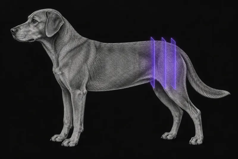
A pelvic fracture is rarely the only injury
Pelvic fractures usually follow major blunt trauma, so the rest of the patient matters as much as the bones:
- Urinary tract. In 585 cats and dogs after vehicular trauma or a fall (Godart et al., New Zealand Veterinary Journal, 2025), lower urinary tract rupture occurred in 3 of 246 dogs (1.2%). Animals with pelvic fractures were 6.4 times more likely to have a rupture. Three of the eight ruptures had no orthopaedic injury, and four of the eight animals could still urinate. All affected animals had free abdominal fluid on abdominal FAST.
- Thorax. In 59 dogs hit by a vehicle (Dancer et al., Veterinary Radiology & Ultrasound, 2019), the sensitivity of thoracic radiographs relative to CT was 69% for lung contusions, 56% for rib fractures, 19% (three views) or 63% (horizontal beam) for pneumothorax, and 43% or 71% for pleural effusion.
When the patient is stable enough for CT, it is worth discussing whether the thorax and abdomen should be scanned in the same session as the pelvis.
Radiographs or CT for the pelvis itself
High-quality radiographs are recommended for every pelvic fracture (Draffan et al., Veterinary and Comparative Orthopaedics and Traumatology, 2009). The question is when CT adds enough to change the plan.
| Study | What it found |
|---|---|
| 13 dogs, two-view and inlet/outlet radiographs vs CT (Crawford et al., Veterinary Radiology & Ultrasound, 2003) | 81% of skeletal lesions seen on CT were diagnosed definitively on radiographs. CT found more abnormalities of the soft tissues, sacroiliac joints and acetabula. All clinically significant surgical lesions were described accurately on radiographs. |
| 25 dogs and cats, orthogonal radiographs vs CT, three orthopaedic surgeons | Management plans did not differ significantly between the two. Disagreement was greatest for complex fractures such as acetabular and sacral fractures, where CT was most sensitive. Experienced surgeons often disagreed with each other. |
| 11 cadavers with induced fractures, four-view radiographs vs CT (Stieger-Vanegas et al., Veterinary and Comparative Orthopaedics and Traumatology, 2015) | Fractures of the sacral body, ischiatic table and pubis were diagnosed more accurately on CT. Acetabular and iliac body fractures were diagnosed with similar accuracy (at least 86%) on either. In trained readers, CT accuracy was close to 100%. |
The authors’ conclusions point the same way. Routine CT may not be justified for diagnosing every pelvic fracture, but it may be advantageous for surgical planning, especially when acetabular fractures are suspected on radiographs. It may also be beneficial where there is uncertainty, particularly with acetabular fractures or sacral fractures that cause neurological deficits.
flowchart TD
  accTitle: Does this dog need pelvic CT after radiographs?
  accDescr: Based on two clinical studies. High-quality radiographs come first for every pelvic fracture. CT is most useful for surgical planning when an acetabular fracture is suspected, a sacral fracture causes neurological deficits, or the radiographs leave uncertainty.
  rads["High-quality pelvic radiographs"] --> q{{"Acetabular, sacral with deficits, or unclear?"}}
  q -- "Yes" --> ct["CT for surgical planning"]
q -- "No" --> plan["Radiographs may be enough"]After surgery: checking implant position
CT multiplanar reconstructions can measure sacroiliac screw angles and pilot hole length relative to sacral width. In a cadaver study of experimental sacroiliac luxations (Déjardin et al., Veterinary and Comparative Orthopaedics and Traumatology, 2016), pilot holes exited the first sacral end-plate in 3 of 10 open-reduction specimens, while the spinal canal was not violated in either surgical group. This was an experimental study, not a clinical series.
Pelvic CT protocol at a glance
General anaesthesia is not always needed. In 14 awake dogs with pelvic trauma (Lee et al., Veterinary Radiology & Ultrasound, 2012), physical restraint was adequate, image quality was good in 10 and fair in 4, and no complications related to restraint were seen. CT also identified more pelvic fractures than radiographs, which overdiagnosed fractures in a few dogs. The images are then read as 2D slices with 3D reconstructions added, not 3D alone (Stieger-Vanegas et al., Veterinary Surgery, 2015).
What the radiologist looks at
- 2D first, 3D added. In a study of 12 dogs with pelvic fractures and 11 cadavers, 3D images were faster to evaluate than 2D images for every reader group. For radiologists and surgeons, though, 2D images were more accurate. 3D improved diagnosis when added to 2D, and reduced accuracy when used without the 2D images.
- Where CT adds most. The soft tissues, sacroiliac joints and acetabula, and the sacral body, ischiatic table and pubis.
- Planning views. Multiplanar and 3D reconstructions were useful for surgical planning in 7 of 13 dogs.
- The thorax, when scanned. Lung contusions, rib fractures, pneumothorax and pleural effusion are more often seen on CT than on radiographs.
What your report will include
Every Veheri case is read by a radiologist or neurologist, and when you order you can choose Diplomate-only reporting or the full specialist panel. You can chat directly with the specialist assigned to your case, and a simplified pet owner report helps you explain the findings to the owner.
When you take the report forward:
- Acetabular or sacral fractures are where CT is most likely to change the description, so share the report with the surgeon before planning.
- The urinary tract still needs checking. A rupture was 6.4 times more likely with pelvic fractures, and an animal that can urinate can still have one.
- New neurological deficits after stabilisation may justify a post-operative CT. This is worth discussing with the surgeon, as the screw position data come from cadavers.
What to include when you request a pelvic CT report
- signalment: age, breed and sex
- mechanism and time of injury
- neurological examination of the pelvic limbs, tail and perineum, including urination and defecation
- the radiographs already taken, and the specific question (for example acetabular involvement or sacral fracture)
- FAST findings and any concern for urinary tract or thoracic injury
- whether the study is pre-operative planning or a post-operative check
How Veheri can help
Veheri’s radiologists report veterinary CT studies remotely for clinics. For other trauma patients, see CT of the canine skull after head trauma and the role of emergency CT.
Sources
Each claim in this guide links to its source. The full citations:
- Godart B, Bonnel G, Bedu AS, et al. Lower urinary tract rupture in cats and dogs following severe blunt trauma. N Z Vet J. 2025;73(1):60-66. doi:10.1080/00480169.2024.2398020
- Dancer SC, Le Roux C, Fosgate GT, et al. Radiography is less sensitive relative to CT for detecting thoracic radiographic changes in dogs affected by blunt trauma secondary to a motor vehicle accident. Vet Radiol Ultrasound. 2019;60(6):648-658. doi:10.1111/vru.12795
- Draffan D, Clements D, Farrell M, et al. The role of computed tomography in the classification and management of pelvic fractures. Vet Comp Orthop Traumatol. 2009;22(3):190-7. doi:10.3415/VCOT-08-04-0035
- Crawford JT, Manley PA, Adams WM. Comparison of computed tomography, tangential view radiography, and conventional radiography in evaluation of canine pelvic trauma. Vet Radiol Ultrasound. 2003;44(6):619-28. doi:10.1111/j.1740-8261.2003.tb00520.x
- Stieger-Vanegas SM, Senthirajah SK, Nemanic S, et al. Evaluation of the diagnostic accuracy of four-view radiography and conventional computed tomography analysing sacral and pelvic fractures in dogs. Vet Comp Orthop Traumatol. 2015;28(3):155-63. doi:10.3415/VCOT-14-06-0096
- Stieger-Vanegas SM, Senthirajah SK, Nemanic S, et al. Evaluation of the diagnostic accuracy of conventional 2-dimensional and 3-dimensional computed tomography for assessing canine sacral and pelvic fractures by radiologists, orthopedic surgeons, and veterinary medical students. Vet Surg. 2015;44(6):694-703. doi:10.1111/j.1532-950X.2014.12313.x
- Lee K, Heng HG, Jeong J, et al. Feasibility of computed tomography in awake dogs with traumatic pelvic fracture. Vet Radiol Ultrasound. 2012;53(4):412-6. doi:10.1111/j.1740-8261.2012.01939.x
- Déjardin LM, Marturello DM, Guiot LP, et al. Comparison of open reduction versus minimally invasive surgical approaches on screw position in canine sacroiliac lag-screw fixation. Vet Comp Orthop Traumatol. 2016;29(4):290-7. doi:10.3415/VCOT-16-02-0030
Related reading
More imaging guides
All imaging guides
CT of adrenal masses in dogs
What contrast CT shows in a dog with an adrenal mass, from caval invasion and adhesions to tumour type, and where its accuracy runs out before adrenalectomy.

Brain CT in dogs when MRI is not available
What brain CT can and cannot detect in dogs when MRI is not available, for masses, stroke, inflammation and head trauma, and when MRI or CSF is still needed.
CT of the canine elbow for medial coronoid disease
When CT is worth it for medial coronoid disease in dogs, what it finds that radiographs miss, and where arthroscopy still gives the better answer.