
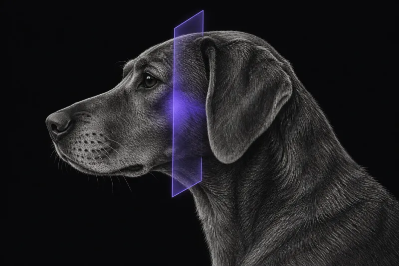
What pituitary imaging is for
Pituitary-dependent hypercortisolism (PDH) is diagnosed with the history, physical examination and endocrine tests, not with MRI. The ACVIM consensus statement (Behrend et al., Journal of Veterinary Internal Medicine, 2013):
- advises endocrine testing only when clinical signs consistent with hypercortisolism are present
- notes that none of the screening or differentiating tests is perfect
- states that imaging can also play a role
Once PDH is confirmed, pituitary MRI answers a different question: how large is the tumour, and where does it extend? That matters because clinical signs can also come from a mass effect of the pituitary tumour on the brain (van Rijn et al., Journal of Veterinary Internal Medicine, 2016), and because size and extension shape the choice between medical treatment, hypophysectomy and radiotherapy.
Which dogs to image
Advanced imaging is not feasible for every dog with hypercortisolism (Yoshida et al., Journal of Veterinary Internal Medicine, 2025). In that study of 130 dogs with hypercortisolism, a pituitary macrotumour was defined as a pituitary height to brain area (P/B) ratio of 0.4 or more. Risk factors were:
- diagnosis at 10.9 years of age or younger
- French Bulldog breed (odds ratio 21.0)
- neurological signs (odds ratio 10.9)
The authors recommend advanced pituitary imaging for dogs with these features. Imaging is also usually needed before transsphenoidal surgery, because one surgical group judges the indication for surgery on an MRI-based classification (Sato et al., Journal of Small Animal Practice, 2016).
flowchart TD
  accTitle: Which dogs with PDH need pituitary imaging?
  accDescr: Based on a 130-dog risk factor study and an MRI-based surgical classification. Dogs with a risk factor for a pituitary macrotumour are recommended for advanced pituitary imaging, and imaging is usually needed before transsphenoidal surgery.
  start["PDH confirmed by endocrine tests"] --> risk{{"Macrotumour risk factor?"}}
  risk -- "Yes" --> mri["Advanced pituitary imaging"]
  risk -- "No" --> surgery{{"Hypophysectomy considered?"}}
surgery -- "Yes" --> mriThe pituitary-to-brain ratio
The P/B ratio relates the height of the pituitary gland to the area of the brain on the same image. In a series of 306 dogs treated by hypophysectomy, a gland was classed as enlarged when the ratio was above 0.31.
| P/B ratio and outcome after hypophysectomy (306 dogs) | Finding |
|---|---|
| Median P/B ratio | 0.39 (range 0.13 to 1.40) |
| Enlarged glands (P/B above 0.31) | 201 dogs |
| Dogs that died within 4 weeks of surgery | Median P/B 0.54, against 0.38 in dogs alive at 4 weeks |
| Survival with P/B above 0.31 | Significantly shorter (hazard ratio 2.34) |
| Disease-free interval with P/B above 0.31 | Significantly shorter (hazard ratio 2.40) |
| Recurrence | 27% of dogs in remission, with a significantly higher P/B ratio than dogs without recurrence |
The authors call the P/B ratio an important pre-operative prognostic factor. They also found that pituitary size at surgery increased over the 20-year study period, while surgery remained an option for larger tumours.
Radiotherapy outcomes
- In 94 dogs treated with radiotherapy for pituitary macroadenomas (Rapastella et al., Journal of Veterinary Internal Medicine, 2023), survival did not differ significantly between dogs with and without PDH. Total radiation dose was the only factor associated with survival on multivariable analysis.
- In 45 dogs treated with stereotactic radiotherapy (Hansen et al., Veterinary Radiology & Ultrasound), dogs with hyperadrenocorticism had a median overall survival of 245 days, against 626 days for dogs with non-functional masses.
Pituitary MRI protocol at a glance
Pituitary size can be measured on post-contrast CT or MRI. In 11 dogs with histologically confirmed adenomas (Auriemma et al., Journal of the American Veterinary Medical Association, 2009), low-field MRI and dynamic CT found an abnormal pituitary in the same 7 dogs, and the P/B ratio did not differ significantly between post-contrast CT and MRI. Signal-to-noise and contrast-to-noise ratios were higher on MRI.
What the radiologist looks at
- Height, not only “enlarged”. The P/B ratio, read against the thresholds used in the hypophysectomy series (above 0.31) and the macrotumour study (0.4 or more).
- Dorsal and craniocaudal extension. The MRI-based classification graded 37 dogs from 1 to 5 by dorsal and craniocaudal extension.
- The surrounding vessels. The same system adds type A or B, depending on whether the arterial circle of Willis or the cavernous sinus was involved. Dogs with type A, grade 1 to 3 tumours had a good prognosis after transsphenoidal hypophysectomy. Grade 3B, 4 and 5 cases may not be suitable for this surgery.
- Resectability. In 34 dogs under 15 kg treated by transsphenoidal surgery (Tanaka et al., Canadian Veterinary Journal, 2025), a higher P/B ratio was associated with partial rather than complete resection.
What your report will include
Every Veheri case is read by a radiologist or neurologist, and when you order you can choose Diplomate-only reporting or the full specialist panel. Neurology reporting covers CT and MRI. You can chat directly with the specialist assigned to your case, and a simplified pet owner report helps you explain the findings to the owner.
When you take the report forward:
- A normal-looking pituitary does not rule out PDH. In the 11-dog comparison, 4 dogs had an adenoma that neither CT nor MRI showed as abnormal.
- Medically managed dogs with a non-enlarged pituitary: the hypophysectomy authors advise yearly re-evaluation including pituitary CT to monitor tumour size.
- Neurological signs that return after radiotherapy may not mean regrowth. In 9 dogs with PDH followed with repeated MRI (Sawada et al., Research in Veterinary Science, 2018), the P/B ratio decreased in all dogs after radiotherapy, and hormone concentrations did not change significantly. In dogs whose neurological signs recurred, MRI showed pituitary haemorrhage rather than tumour regrowth.
- Talking to the owner: after hypophysectomy, a P/B ratio above 0.31 came with significantly shorter survival and disease-free interval, so tumour size belongs in the treatment discussion.
What to include when you request a pituitary MRI report
- signalment: age, breed and sex
- the endocrine tests that confirmed PDH and their dates
- any neurological signs
- current treatment and how long it has been given
- whether hypophysectomy or radiotherapy is being considered
- previous pituitary imaging, so size can be compared
How Veheri can help
Veheri provides veterinary teleradiology and teleneurology for X-ray, CT and MRI, with expert support for MRI scanning protocols, positioning and acquisition. Related reading: pituitary imaging for hypersomatotropism in cats and when veterinary MRI is the right move.
Sources
Each claim in this guide links to its source. The full citations:
- Behrend EN, Kooistra HS, Nelson R, et al. Diagnosis of spontaneous canine hyperadrenocorticism: 2012 ACVIM consensus statement (small animal). J Vet Intern Med. 2013;27(6):1292-304. doi:10.1111/jvim.12192
- van Rijn SJ, Galac S, Tryfonidou MA, et al. The influence of pituitary size on outcome after transsphenoidal hypophysectomy in a large cohort of dogs with pituitary-dependent hypercortisolism. J Vet Intern Med. 2016;30(4):989-95. doi:10.1111/jvim.14367
- Sato A, Teshima T, Ishino H, et al. A magnetic resonance imaging-based classification system for indication of trans-sphenoidal hypophysectomy in canine pituitary-dependent hypercortisolism. J Small Anim Pract. 2016;57(5):240-6. doi:10.1111/jsap.12474
- Yoshida K, Kobatake Y, Takashima S, et al. Risk factors for pituitary macrotumor in dogs with hypercortisolism. J Vet Intern Med. 2025;39(6):e70261. doi:10.1111/jvim.70261
- Tanaka S, Suzuki S, Teshima T, et al. Outcomes of transsphenoidal surgery in 34 small dogs with pituitary-dependent hypercortisolism. Can Vet J. 2025;66(10):1094-1103. PMID: 41030423. https://pubmed.ncbi.nlm.nih.gov/41030423/
- Auriemma E, Barthez PY, van der Vlugt-Meijer RH, et al. Computed tomography and low-field magnetic resonance imaging of the pituitary gland in dogs with pituitary-dependent hyperadrenocorticism: 11 cases (2001-2003). J Am Vet Med Assoc. 2009;235(4):409-14. doi:10.2460/javma.235.4.409
- Rapastella S, Morabito S, Sharman M, et al. Effect of pituitary-dependent hypercortisolism on the survival of dogs treated with radiotherapy for pituitary macroadenomas. J Vet Intern Med. 2023;37(4):1331-1340. doi:10.1111/jvim.16724
- Hansen KS, Zwingenberger AL, Théon AP, et al. Long-term survival with stereotactic radiotherapy for imaging-diagnosed pituitary tumors in dogs. Vet Radiol Ultrasound. 60(2):219-232 (published online 2018). doi:10.1111/vru.12708
- Sawada H, Mori A, Lee P, et al. Pituitary size alteration and adverse effects of radiation therapy performed in 9 dogs with pituitary-dependent hypercortisolism. Res Vet Sci. 2018;118:19-26. doi:10.1016/j.rvsc.2018.01.001
Related reading
More imaging guides
All imaging guides
Pituitary imaging for hypersomatotropism in cats
When diabetic cats need pituitary imaging for hypersomatotropism, what MRI and CT show, and why a normal-sized gland does not rule it out.
MRI of the canine brachial plexus for peripheral nerve sheath tumours
How MRI finds peripheral nerve sheath tumours of the canine brachial plexus, what the radiologist looks for, and what imaging can say about grade and extent.
Brain MRI for dogs with seizures
Which dogs with seizures need brain MRI, what the evidence says about diagnostic yield, what the radiologist looks for and what your report will include.