
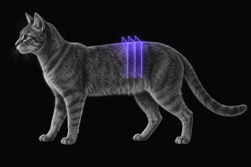
Disc disease in cats is uncommon, but real
- Prevalence. Of 12,900 cats seen at one referral hospital (De Decker et al., Journal of Feline Medicine and Surgery, 2017), 31 (0.24%) were diagnosed with thoracolumbar intervertebral disc disease. Purebred cats, British Shorthairs (1.29% of those presented) and Persians (1.83%) were overrepresented.
- Incidence. In two other referral populations (Amey et al., Journal of Veterinary Internal Medicine, 2024), the incidence of thoracolumbar and lumbosacral disc herniation was 0.44% (92 of 20,849 cats).
- Location. A systematic review of 93 cats with analysable data (Ebeling et al., Journal of Feline Medicine and Surgery, 2025) found lumbar involvement in 59%, thoracolumbar in 31% and cervical in 5%.
Degeneration is common and often incidental
- Asymptomatic cats. In T2-weighted MRI of 60 cadavers of cats without spinal signs (Schmid et al., American Journal of Veterinary Research, 2024), 1,544 discs were graded. Of these, 57.3% were Pfirrmann grade 1, 27.5% grade 2, 9.5% grade 3, 5.3% grade 4 and 0.4% grade 5. Middle-aged (odds ratio 4.03) and old cats (12.5) had higher odds of degeneration than young cats.
- Cats with myelopathy. In 114 cats with suspected myelopathy that had spinal MRI (Soteras et al., Journal of the American Veterinary Medical Association, 2024), 70% had at least one degenerative change. Degenerative lumbosacral stenosis was commonly considered incidental.
The key reporting question is therefore not “is there a disc lesion?” but “does this lesion explain the neurological findings?”
What the evidence says about outcome
- Surgery and conservative care. In the 92 cats with compressive herniation between T3 and S1, success was 62% at 6 weeks and 74% at 6 months after surgery, and 54% and 65% after conservative treatment. Cats with a history of trauma were more likely to do well at 6 weeks (odds ratio 5.3). Neurological deficits persisted in most cats at 6 weeks (86% surgical, 92% conservative).
- Pooled data. Across 93 cats in the systematic review, 85% had a positive outcome, but no statistically significant prognostic factor was found. The authors note the low level of evidence.
- Non-compressive lesions. In 11 cats with presumptive acute non-compressive nucleus pulposus extrusion (Taylor-Brown and De Decker, Journal of Feline Medicine and Surgery, 2017), treatment included supportive care, and all cats that presented non-ambulatory regained ambulation. Most of 12 cats with presumptive intramedullary extrusion (Heaselgrave et al., Journal of Feline Medicine and Surgery, 2025) had a favourable outcome.
Feline spinal MRI protocol at a glance
Because the lumbar spine dominates, it is usually worth including the whole lumbar column, not only the thoracolumbar junction, when signs allow a T3–S3 localisation. The study also has to separate compressive, non-compressive and intramedullary disc lesions, since the last two are presumptive MRI diagnoses.
What the radiologist looks at
In 43 cats with 50 disc herniations (Bibbiani et al., Journal of Feline Medicine and Surgery, 2022), the types were acute non-compressive nucleus pulposus extrusion (ANNPE, 22), disc protrusion (19) and disc extrusion (9). Localisation was L4–S3 in 19 cats, T3–L3 in 18 and C1–C5 in 6. The radiologist decides which type is present and whether it fits the signs:
| Type | Clinical pattern reported | What MRI must show or decide |
|---|---|---|
| Disc extrusion | Shorter duration and more severe deficits than protrusion (12,900-cat study); associated with spinal hyperaesthesia and males (43-cat study) | Compressive extradural material. When present, it was always responsible for the signs in one series |
| Disc protrusion | Longer duration, milder deficits, more often multiple (12,900-cat and 43-cat studies) | Whether it explains the signs; it was infrequently related to them |
| ANNPE | Single site, more often females, not associated with spinal pain (43-cat study); peracute, often after trauma (11-cat study) | By definition, no significant cord compression, and treatment in one series of 11 cats included supportive care |
| Intramedullary extrusion | Peracute or acute, with confirmed or possible trauma in 9 of 12 cats; sites from T12–T13 to L5–L6 (12-cat study) | Disc material within the cord parenchyma |
The type is not always clear cut. Acute-on-chronic herniations with features of both extrusion and protrusion were found in 10% of cats in one outcome study.
A differential for the chronic T3–L3 cat. In 9 cats with thoracic vertebral canal stenosis (Gillespie and De Decker, Journal of Feline Medicine and Surgery, 2020), all had a chronic, progressive, painful, ambulatory T3–L3 myelopathy. British Shorthairs and male neutered cats were overrepresented and the median age was 9 years. Affected cats had a lower vertebral canal height than controls at several thoracic levels. Canal narrowing is worth assessing explicitly in British Shorthairs whose discs do not explain the signs.
What your report will include
Every Veheri case is read by a radiologist or neurologist, and when you order you can choose Diplomate-only reporting or the full specialist panel. Neurology reporting covers CT and MRI. You can chat directly with the specialist assigned to your case, and a simplified pet owner report helps you explain the findings to the owner.
When you take the report forward:
- Read degenerative findings against the signs. With degeneration common in cats without spinal signs, a degenerate disc is not automatically the cause.
- ANNPE and intramedullary extrusion are presumptive MRI diagnoses, as in the 11-cat and 12-cat series.
- Talking to the owner: MRI type and location have not been shown to predict outcome in cats, and neurological deficits persisted in most cats at 6 weeks whichever treatment was chosen, so it helps to set expectations early.
What to include when you request a spinal MRI report
- signalment: age, breed and sex
- onset, duration and progression, and whether the cat is painful
- any trauma before onset
- breed, especially British Shorthair or Persian
- ambulatory status, nociception and continence
- previous episodes, treatment or spinal surgery
How Veheri can help
Veheri provides veterinary teleradiology and teleneurology and reports spinal MRI studies remotely for clinics. For other causes of myelopathy in cats, see spinal cord MRI in cats, and for dogs, MRI of the canine thoracolumbar spine for disc extrusion.
Sources
Each claim in this guide links to its source. The full citations:
- De Decker S, Warner AS, Volk HA. Prevalence and breed predisposition for thoracolumbar intervertebral disc disease in cats. J Feline Med Surg. 2017;19(4):419-423. doi:10.1177/1098612X16630358
- Amey JA, Liatis T, Cherubini GB, et al. Outcomes of surgically and conservatively managed thoracolumbar and lumbosacral intervertebral disc herniations in cats. J Vet Intern Med. 2024;38(1):247-257. doi:10.1111/jvim.16950
- Ebeling R, Lorenz N, Zablotski Y, et al. Feline intervertebral disc disease: a systematic review and meta-analysis. J Feline Med Surg. 2025;27(12):1098612X251385878. doi:10.1177/1098612X251385878
- Bibbiani L, Gelendi S, Bernardini M, et al. Prevalence, clinical presentation and MRI of intervertebral disc herniations in cats. J Feline Med Surg. 2022;24(12):e443-e452. doi:10.1177/1098612X221121893
- Soteras MP, Dominguez E, Suñol A, et al. Spinal magnetic resonance imaging in cats: differences in clinical significance of intervertebral disk extrusion, intervertebral disk protrusion, and degenerative lumbosacral stenosis. J Am Vet Med Assoc. 2024;262(9):1193-1200. doi:10.2460/javma.24.02.0099
- Taylor-Brown FE, De Decker S. Presumptive acute non-compressive nucleus pulposus extrusion in 11 cats: clinical features, diagnostic imaging findings, treatment and outcome. J Feline Med Surg. 2017;19(1):21-26. doi:10.1177/1098612X15605150
- Heaselgrave T, De Risio L, Domínguez E, et al. Presumptive intramedullary intervertebral disc extrusion in 12 cats: clinical presentation, MRI findings, treatment and outcome. J Feline Med Surg. 2025;27(7):1098612X251349712. doi:10.1177/1098612X251349712
- Schmid D, Lanzillotta V, Evans R, et al. The prevalence of intervertebral disc degeneration in the cervical, thoracic, and lumbar spine in asymptomatic cats. Am J Vet Res. 2024;85(12). doi:10.2460/ajvr.24.04.0095
- Gillespie S, De Decker S. Thoracic vertebral canal stenosis in cats: clinical features, diagnostic imaging findings, treatment and outcome. J Feline Med Surg. 2020;22(12):1191-1199. doi:10.1177/1098612X20920041
Related reading
More imaging guides
All imaging guides
CT of the canine thoracolumbar spine for suspected disc extrusion
When non-contrast CT is enough for thoracolumbar disc extrusion in chondrodystrophic dogs, and which dogs need CT-myelography or MRI instead.
MRI of the canine thoracolumbar spine for disc extrusion
What MRI shows in dogs with acute thoracolumbar disc extrusion, how it compares with CT, and which findings matter for surgery, prognosis and myelomalacia.
Brain MRI in cats
What brain MRI shows in cats with seizures, meningioma, lymphoma or FIP, how often the scan is normal, and what MRI cannot settle on its own.