
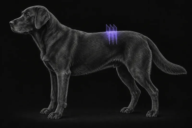
Why CT works well in chondrodystrophic dogs
In chondrodystrophic dogs, extruded disc material is often mineralised, and plain CT enables the diagnosis in most cases (Robertson and Thrall, Veterinary Radiology & Ultrasound, 2011). In 46 dogs with acute myelopathy (Dennison et al., Veterinary Radiology & Ultrasound, 2010), 21 of 23 Hansen type I disc extrusions were mineralised, but none of the Hansen type II protrusions were.
CT is also more widely available than MRI, less costly and much faster, and it can be performed under sedation, as a 2020 review of imaging in disc disease (da Costa et al., Frontiers in Veterinary Science) notes. In 19 chondrodystrophic dogs with acute thoracolumbar extrusion (Hecht et al., Veterinary Radiology & Ultrasound, 2009), the level of extrusion agreed with surgery in 100% of dogs on conventional CT, 94.7% on helical CT and 94.7% on myelography. Median examination times were 4 minutes for helical CT, 8 for conventional CT and 32 for myelography.
One outcome study supports this. In 560 non-ambulatory chondrodystrophic dogs with acute thoracolumbar extrusion (Low and Markovic, Veterinary Surgery, 2026), management guided by conditional CT-myelography (42 dogs) was non-inferior to MRI (518 dogs) for recovery of ambulation. Rates of postoperative deterioration, progressive myelomalacia and major complications did not differ, and costs were lower.
How CT compares with MRI
MRI remains the more sensitive test.
- Detection. In 44 dogs with surgically confirmed disc herniation (Cooper et al., Veterinary Radiology & Ultrasound, 2014), non-contrast CT had a sensitivity of 88.6% and MRI 98.5%. MRI was more accurate for the site of compression and for separating extrusion from protrusion.
- Who CT misses. In that study, CT was less accurate for localisation in peracute cases and in chondrodystrophic, female, older and smaller dogs (under 7 kg). In 182 dogs (Israel et al., Veterinary Radiology & Ultrasound, 2009), myelography was more sensitive than CT in dogs under 5 kg, while CT was more sensitive in chronically affected dogs.
- Surgical plan. When four observers planned hemilaminectomies in 40 chondrodystrophic dogs (Noyes et al., Veterinary Surgery, 2017), plans based on CT and MRI of the same dog were similar in 43.5% to 66.6% of dogs. The side was the most consistent element; observers tended to plan larger defects on MRI.
When CT is not enough
Non-contrast CT shows mineralised disc material and loss of epidural fat, but not the spinal cord itself, and a disc extrusion can go undetected on CT. The evidence supports further imaging, with CT-myelography or MRI, in these situations:
| Situation | Evidence |
|---|---|
| No lesion on CT, or a lesion that does not match the neurolocalisation | Further imaging may be required; CT-myelography was often necessary when CT showed no lesion |
| Non-chondrodystrophic dog | Additional imaging was needed in 17.4% of non-chondrodystrophic breeds versus 3.6% of Dachshunds in a study of 555 dogs; CT-myelography was often necessary in these dogs |
| Plegic dog | Concurrent extradural compression and spinal cord swelling can require CT-myelography for a correct diagnosis |
| Very small dog | CT was less accurate under 7 kg and less sensitive than myelography under 5 kg |
| Suspected non-compressive or hydrated extrusion | MRI is the modality of choice for acute non-compressive and hydrated nucleus pulposus extrusion |
Thoracolumbar spine CT protocol at a glance
Because extruded disc material in chondrodystrophic dogs is often mineralised, plain CT enables the diagnosis in most cases. Multiplanar reconstructions add confidence: in 111 dogs they increased mean certainty scores for a correct diagnosis by 3–11% compared with 2D CT images. When more is needed, CT-myelography was the most sensitive technique for compression in 46 dogs with acute myelopathy, with the highest agreement between observers.
What the radiologist looks at
The 2020 review summarises a series of 23 surgically confirmed thoracolumbar extrusions that fell into three CT patterns:
| Pattern | CT appearance |
|---|---|
| Acutely extruded, mineralised nucleus pulposus | Heterogeneous, hyperattenuating extradural mass (mean 219 HU) with severe compression |
| Acute extrusion with haemorrhage | Less compression, extending over several vertebrae, only slightly distinguishable from the cord (mean 59 HU) |
| Chronic mineralised nucleus pulposus | Very hyperattenuating, more homogeneous material (mean 745 HU) |
- The haemorrhagic pattern deserves particular care, because its attenuation is close to that of the cord.
- Does the lesion match? The radiologist checks the lesion against the neuroanatomical localisation you send, because a mismatch is a reason for further imaging.
- Which side, and how far? The side was the most consistent element between CT- and MRI-based plans, but overall plans from CT and MRI of the same dog were similar in only 43.5% to 66.6% of dogs.
What your report will include
Every Veheri case is read by a radiologist or neurologist, and when you order you can choose Diplomate-only reporting or the full specialist panel. Neurology reporting covers CT and MRI. You can chat directly with the specialist assigned to your case, and a simplified pet owner report helps you explain the findings to the owner.
When you take the report forward:
- A negative or mismatched CT is a reason for CT-myelography or MRI, not a normal result, because a disc extrusion can go undetected on CT.
- Prognosis needs the cord. For acute non-compressive and hydrated nucleus pulposus extrusion, MRI findings have been linked to the likelihood of recovery. Where that would change the decision, MRI is worth discussing even when CT has already shown the likely cause.
- Surgical planning: observers tended to plan larger defects on MRI than on CT, so discuss the extent of the approach with the surgeon.
- Talking to the owner: in non-ambulatory chondrodystrophic dogs, management guided by conditional CT-myelography was non-inferior to MRI for recovery of ambulation, and costs were lower.
What to include when you request a spinal CT report
- signalment: age, breed and sex
- body weight, since CT was less accurate in small dogs
- onset and duration of signs, since peracute and chronic cases behave differently on CT
- neurological grade, including ambulation and nociception
- neuroanatomical localisation, so the radiologist can check the lesion matches
- previous spinal imaging or surgery
- whether CT-myelography or MRI is available if CT is inconclusive
How Veheri can help
Veheri works with veterinary radiologists and neurologists and reports CT and MRI studies remotely for clinics. For the MRI side of disc disease, see MRI of the thoracolumbar spine in dogs, and for scanning, see mastering veterinary spinal CT.
Sources
Each claim in this guide links to its source. The full citations:
- Robertson I, Thrall DE. Imaging dogs with suspected disc herniation: pros and cons of myelography, computed tomography, and magnetic resonance. Vet Radiol Ultrasound. 2011;52(1 Suppl 1):S81-4. doi:10.1111/j.1740-8261.2010.01788.x
- Dennison SE, Drees R, Rylander H, et al. Evaluation of different computed tomography techniques and myelography for the diagnosis of acute canine myelopathy. Vet Radiol Ultrasound. 2010;51(3):254-8. doi:10.1111/j.1740-8261.2010.01667.x
- da Costa RC, De Decker S, Lewis MJ, et al. Diagnostic imaging in intervertebral disc disease. Front Vet Sci. 2020;7:588338. doi:10.3389/fvets.2020.588338
- Hecht S, Thomas WB, Marioni-Henry K, et al. Myelography vs. computed tomography in the evaluation of acute thoracolumbar intervertebral disk extrusion in chondrodystrophic dogs. Vet Radiol Ultrasound. 2009;50(4):353-9. doi:10.1111/j.1740-8261.2009.01549.x
- Low D, Markovic A. Computed tomography versus magnetic resonance imaging for the perioperative management of acute thoracolumbar intervertebral disc extrusion in non-ambulatory chondrodystrophic dogs. Vet Surg. 2026;55(7):1198-1211. doi:10.1111/vsu.70135
- Cooper JJ, Young BD, Griffin JF, et al. Comparison between noncontrast computed tomography and magnetic resonance imaging for detection and characterization of thoracolumbar myelopathy caused by intervertebral disk herniation in dogs. Vet Radiol Ultrasound. 2014;55(2):182-9. doi:10.1111/vru.12114
- Israel SK, Levine JM, Kerwin SC, et al. The relative sensitivity of computed tomography and myelography for identification of thoracolumbar intervertebral disk herniations in dogs. Vet Radiol Ultrasound. 2009;50(3):247-52. doi:10.1111/j.1740-8261.2009.01528.x
- Noyes JA, Thomovsky SA, Chen AV, et al. Magnetic resonance imaging versus computed tomography to plan hemilaminectomies in chondrodystrophic dogs with intervertebral disc extrusion. Vet Surg. 2017;46(7):1025-1031. doi:10.1111/vsu.12700
Related reading
More imaging guides
All imaging guides
MRI of the canine thoracolumbar spine for disc extrusion
What MRI shows in dogs with acute thoracolumbar disc extrusion, how it compares with CT, and which findings matter for surgery, prognosis and myelomalacia.
MRI of the feline thoracolumbar spine for disc disease
How MRI classifies disc extrusion, protrusion and non-compressive extrusion in cats, why degeneration is often incidental, and what outcome data show.

CT of adrenal masses in dogs
What contrast CT shows in a dog with an adrenal mass, from caval invasion and adhesions to tumour type, and where its accuracy runs out before adrenalectomy.