
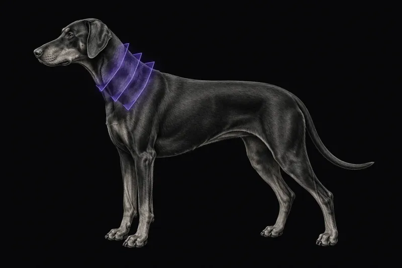
Two forms, often in different dogs
Cervical spondylomyelopathy (CSM), or wobbler syndrome, has a disc-associated form (DA-CSM) and an osseous-associated form (OA-CSM). MRI is usually used to characterise the form and to show each site of compression.
- Disc-associated CSM is the most common form. Compression is typically caused by protrusion of one or more caudal cervical discs, sometimes with dorsal compression from ligamentum flavum hypertrophy. In a review of DA-CSM (De Decker et al., Veterinary Record, 2012), the C6-C7 disc space is most often affected, and adult to older Dobermanns are over-represented. In 63 dogs on high-field MRI (Bonelli et al., BMC Veterinary Research, 2021), the median age was 7.2 years and the main compression was commonly at C6-C7 or C5-C6.
- Osseous-associated CSM affects younger dogs. In 100 dogs on high-field MRI (Bonelli et al., Veterinary Radiology & Ultrasound, 2021), 73% were giant breeds and 27% large breeds, with a median age of 2 years.
- Both forms together are common. Of 232 dogs with CSM (Bonelli and da Costa, Journal of Veterinary Internal Medicine, 2023), 60 had disc protrusion and osseous proliferation. In 40 of these 60 dogs, both types of compression were at the same site, and those dogs were more likely to have a higher neurological grade.
What MRI shows
| Finding | Disc-associated CSM (63 dogs) | Osseous-associated CSM (100 dogs) |
|---|---|---|
| More than one site of spinal cord compression | 57% | 78% |
| Foraminal stenosis | 81% | 91% |
| Other changes | T2-weighted spinal cord hyperintensity in 63%; ligamentum flavum hypertrophy in 38%; degenerative changes in 88% of articular processes | Intervertebral disc degeneration in 80%; ligamentum flavum or soft tissue proliferation more likely with multiple sites |
Most dogs in both series had a chronic presentation (83% and 89%).
Traction and flexion-extension studies
Compression in CSM can change with neck position. Two techniques address this.
Traction MRI shows whether a disc-associated lesion is traction-responsive (dynamic) or static. In 25 Dobermanns on low-field MRI (Stabile et al., Journal of Small Animal Practice, 2015), neurological grade was not associated with whether the C6-C7 lesion was static or traction-responsive. C7-T1 disc degeneration was associated with a traction-responsive lesion at C6-C7. Traction does not assess flexion and extension.
Kinematic MRI images the neck in neutral, flexed and extended positions:
- In 9 Dobermanns with DA-CSM (Provencher et al., Journal of Veterinary Internal Medicine, 2016), flexion improved or resolved compression in 4 dogs, and extension worsened compression in 6. Extension revealed 6 new compressive lesions.
- In 12 dogs with OA-CSM (Provencher et al., Veterinary Radiology & Ultrasound, 2017), 25 compressions were seen in neutral position and 32 in extension. Extension changed the most severe site of compression in 4 of the 12 dogs. None of the dogs deteriorated neurologically after kinematic imaging.
Both kinematic studies were small, prospective and from one research group.
Cervical spine MRI protocol at a glance
A standard cervical MRI study characterises the form of CSM and shows every site of compression, as in the disc-associated and osseous-associated high-field series. Dynamic studies are optional additions: traction MRI repeats sequences with linear traction on the neck, and kinematic MRI uses a positioning device to image flexion and extension, as described in the DA-CSM kinematic study.
What the radiologist looks at
- Every site, not only the worst. Several sites and foraminal stenosis are common, so each site, the direction of compression and the foramina are described.
- Compression is not the same as disease. MRI abnormalities are found in clinically normal dogs of predisposed breeds. In 15 clinically normal and 15 affected Great Danes (Martin-Vaquero and da Costa, Journal of the American Veterinary Medical Association, 2014), MRI detected 11 compressive sites in the normal dogs and 61 in the affected dogs. Foraminal stenosis was present in 11 of the normal dogs.
- What separates affected dogs. In the same study, spinal cord signal changes were seen only in affected dogs. Severe spinal cord compression, the number of stenotic foraminal sites and spinal cord signal changes distinguished affected from normal dogs.
- The fit with the neurological grade. In DA-CSM, severity of compression correlated moderately with neurological grade (r 0.48), but the number of affected sites did not. In OA-CSM, neurological grade correlated only weakly with compression severity (r = 0.27), number of sites (r = 0.24) and T2-weighted hyperintensity (r = 0.24). The MRI is therefore read together with the neurological examination.
What your report will include
Every Veheri case is read by a radiologist or neurologist, and when you order you can choose Diplomate-only reporting or the full specialist panel. Neurology reporting covers CT and MRI. You can chat directly with the specialist assigned to your case, and a simplified pet owner report helps you explain the findings to the owner.
When you take the report forward:
- Deciding on a dynamic study is usually for the neurologist or surgeon who plans treatment. It matters most when the choice of surgical technique depends on it, because extension changed the most severe site of compression in 4 of 12 dogs.
- Follow-up MRI is interpreted against the clinical course. In 11 dogs with OA-CSM re-imaged after a median of 30 months (Nye et al., Journal of Veterinary Internal Medicine, 2020), the most affected site in medically treated dogs had progressed in 4 of 9 dogs and improved in 4. Most dogs were clinically unchanged or improved.
- Talking to the owner: compression on MRI in a predisposed breed does not by itself prove the cause of the signs, so the neurological examination stays central.
What to include when you request a cervical MRI report
- signalment: age, breed and sex
- body weight
- duration and course of signs: chronic, progressive or acute
- neurological grade, and whether neck pain, thoracic limb signs or pelvic limb ataxia dominate
- whether traction or flexion-extension imaging is wanted, and who will plan treatment
- previous cervical surgery or imaging, so sites can be compared
How Veheri can help
Veheri provides veterinary teleradiology and teleneurology for X-ray, CT and MRI, with expert support for MRI scanning protocols, positioning and acquisition. Related reading: MRI of the craniocervical junction, MRI of the canine thoracolumbar spine and when veterinary MRI is the right move.
Sources
Each claim in this guide links to its source. The full citations:
- Bonelli MA, da Costa LBSBC, da Costa RC. Magnetic resonance imaging and neurological findings in dogs with disc-associated cervical spondylomyelopathy: a case series. BMC Vet Res. 2021;17(1):145. doi:10.1186/s12917-021-02846-5
- Bonelli MA, da Costa LBSBC, da Costa RC. Association of neurologic signs with high-field MRI findings in 100 dogs with osseous-associated cervical spondylomyelopathy. Vet Radiol Ultrasound. 2021;62(6):678-686. doi:10.1111/vru.13014
- De Decker S, da Costa RC, Volk HA, Van Ham LM. Current insights and controversies in the pathogenesis and diagnosis of disc-associated cervical spondylomyelopathy in dogs. Vet Rec. 2012;171(21):531-7. doi:10.1136/vr.e7952
- Bonelli MA, da Costa RC. Magnetic resonance imaging and neurologic characterization of combined osseous- and disc-associated cervical spondylomyelopathy in dogs. J Vet Intern Med. 2023;37(4):1418-1427. doi:10.1111/jvim.16792
- Martin-Vaquero P, da Costa RC. Magnetic resonance imaging features of Great Danes with and without clinical signs of cervical spondylomyelopathy. J Am Vet Med Assoc. 2014;245(4):393-400. doi:10.2460/javma.245.4.393
- Provencher M, Habing A, Moore SA, et al. Kinematic magnetic resonance imaging for evaluation of disc-associated cervical spondylomyelopathy in Doberman Pinschers. J Vet Intern Med. 2016;30(4):1121-8. doi:10.1111/jvim.13981
- Stabile F, Bernardini M, Bevilacqua G, et al. Neurological signs and pre- and post-traction low-field MRI findings in Dobermanns with disc-associated cervical spondylomyelopathy. J Small Anim Pract. 2015;56(5):331-8. doi:10.1111/jsap.12326
- Provencher M, Habing A, Moore SA, et al. Evaluation of osseous-associated cervical spondylomyelopathy in dogs using kinematic magnetic resonance imaging. Vet Radiol Ultrasound. 2017;58(4):411-421. doi:10.1111/vru.12495
- Nye C, Hostnik E, Parker E, et al. Long-term clinical and magnetic resonance imaging follow-up of dogs with osseous-associated cervical spondylomyelopathy. J Vet Intern Med. 2020;34(5):2012-2020. doi:10.1111/jvim.15866
Related reading
More imaging guides
All imaging guides
MRI of the canine brachial plexus for peripheral nerve sheath tumours
How MRI finds peripheral nerve sheath tumours of the canine brachial plexus, what the radiologist looks for, and what imaging can say about grade and extent.
Brain MRI for dogs with seizures
Which dogs with seizures need brain MRI, what the evidence says about diagnostic yield, what the radiologist looks for and what your report will include.
MRI of the craniocervical junction for Chiari-like malformation and syringomyelia
What MRI of the canine craniocervical junction should include for Chiari-like malformation and syringomyelia, and how to read a syrinx against the signs.