
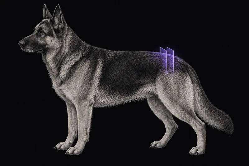
Why lumbosacral MRI needs careful reading
Degenerative lumbosacral stenosis (DLSS) is the most common disorder of the caudal lumbar spine in dogs (Meij and Bergknut, Veterinary Clinics of North America: Small Animal Practice, 2010). Affected dogs are typically “neuro-orthopaedic” patients, and the most consistent sign is lumbosacral pain. CT and MRI show intervertebral disc degeneration, compression of the cauda equina and entrapment of nerve roots, which allows treatment to be tailored to the dog.
The difficulty is that definitive criteria for a reliable diagnosis remain elusive (Jeffery et al., The Veterinary Journal, 2014), whether from examination, imaging or electrodiagnostics. MRI is the most commonly used advanced imaging test for DLSS, but stenosis of the central canal and foramina does not reliably correlate with clinical signs (Siauciunaite et al., The Veterinary Journal, 2026).
Degeneration is common in dogs without signs
- Young German Shepherd Dogs. In 110 clinically sound German Shepherd Dogs and 47 healthy dogs of other large breeds (Amort et al., Veterinary Radiology & Ultrasound, 2012), most younger than 18 months, the lumbosacral disc of German Shepherd Dogs showed significantly higher grades of degeneration.
- Healthy large breed dogs. In 22 healthy large breed dogs imaged in different positions (Lampe et al., Veterinary Radiology & Ultrasound, 2020), subclinical compression was seen in 45% in neutral position and 85% in extension. Disc protrusion was seen in 55% and 73% respectively.
Compression at the lumbosacral junction is therefore common in dogs without signs. On its own it does not prove that DLSS causes the signs.
Findings that relate to clinical signs
In the studies below, foraminal and nerve root findings were the ones associated with clinical signs.
| MRI finding | What the studies found |
|---|---|
| Foraminal stenosis | Present in 66% of 50 dogs with DLSS and in 1 of 17 controls. Unilateral clinical signs were associated with ipsilateral foraminal stenosis (Siauciunaite et al.). |
| Lumbar nerve root changes | Present in 60% of dogs with DLSS and 2 of 17 controls. Ipsilateral thickening of the L7 nerve root was associated with unilateral nerve root dysfunction (Siauciunaite et al.). |
| Loss of foraminal fat signal | In 240 dogs, 480 L7 foramina, on low-field MRI (Lichtenhahn et al., Veterinary Surgery, 2020), 364 foramina had loss of fat signal, and 87.9% of those also had nerve root changes. |
| Unilateral imaging findings | Clinical signs were in the ipsilateral pelvic limb in 85% of dogs with unilateral findings (Lichtenhahn et al.). |
| Nerve root sedimentation sign | Did not differ significantly between dogs with DLSS and controls (Siauciunaite et al.). |
| Redundant nerve roots | Small to moderate association with DLSS (Siauciunaite et al.). |
Position changes the picture
The lumbosacral junction moves, and MRI measurements change with it.
- In the healthy dogs, most measurements were larger in flexion and smaller in extension than in neutral position.
- In 24 clinically affected dogs (Baldo Clemot et al., Veterinary Surgery), disc protrusion, foraminal stenosis and ventral bulging of the ligamentum flavum were significantly reduced in flexion. No dog had persistent cauda equina compression or completely occluded foramina in flexion.
Lumbosacral MRI protocol at a glance
The main protocol choice at the lumbosacral junction is position, because measurements change between neutral, flexed and extended positions. Whether a flexed or extended series is added is usually agreed with the surgeon in advance. For the L7 nerve roots, the low-field foraminal study relied on dorsal oblique and transverse images.
What the radiologist looks at
- The foramina and the side of the signs. Foraminal stenosis and nerve root changes are matched to the side of the clinical signs, because unilateral signs were associated with ipsilateral findings.
- Signs of L7 radiculopathy. On low-field MRI, these included nerve root enlargement and hyperintensity, and changes in nerve root shape, size or position.
- Compression in context. Disc protrusion and compression are common in healthy dogs, so they are weighed against the examination, not read as proof of DLSS.
- Signs that do not discriminate. The nerve root sedimentation sign did not separate dogs with DLSS from controls. In the healthy dogs, agreement between observers was poor for loss of foraminal fat signal and for evidence of compression.
What your report will include
Every Veheri case is read by a radiologist or neurologist, and when you order you can choose Diplomate-only reporting or the full specialist panel. Neurology reporting covers CT and MRI. You can chat directly with the specialist assigned to your case, and a simplified pet owner report helps you explain the findings to the owner.
When you take the report forward:
- Imaging and surgery do not always agree. In 35 dogs (Suwankong et al., Journal of the American Veterinary Medical Association, 2006), CT and MRI agreed closely with each other, for example on the degree of disc protrusion (kappa 0.88). Agreement between either modality and the surgical findings was only moderate for disc protrusion (kappa 0.44 to 0.56) and nerve root swelling (kappa 0.40 and 0.50).
- Extrusion or protrusion. In 117 dogs with surgically confirmed lumbosacral disc herniation (Shing et al., Frontiers in Veterinary Science, 2023), low-field MRI separated extrusion from protrusion with an overall accuracy of 68.8% and fair agreement between radiologists. Accuracy rose to 85.3% for diagnoses made with confidence.
- Surgical technique. The authors of the flexion study suggest that, when distraction-stabilisation is used, the need for added decompression should be questioned, and call for surgical case-control studies.
- Talking to the owner: because definitive diagnostic criteria remain elusive, MRI findings are interpreted together with the examination. Explain that a scan can show changes that are not the cause of the signs.
What to include when you request a lumbosacral MRI report
- signalment: age, breed and sex
- the dog’s activity or working use
- the main sign, such as lumbosacral pain or pelvic limb lameness, and any tail or continence signs
- which side is affected, since unilateral signs can be matched to foraminal and nerve root findings
- orthopaedic findings, because these dogs are often neuro-orthopaedic patients and joint disease may coexist
- whether surgery is planned and which technique, so the radiologist knows whether flexed or extended imaging is useful
How Veheri can help
Veheri works with veterinary radiologists and neurologists and reports spinal MRI studies remotely for clinics. For disc disease further forward, see MRI of the canine thoracolumbar spine, and read when veterinary MRI is the right move.
Sources
Each claim in this guide links to its source. The full citations:
- Meij BP, Bergknut N. Degenerative lumbosacral stenosis in dogs. Vet Clin North Am Small Anim Pract. 2010;40(5):983-1009. doi:10.1016/j.cvsm.2010.05.006
- Jeffery ND, Barker A, Harcourt-Brown T. What progress has been made in the understanding and treatment of degenerative lumbosacral stenosis in dogs during the past 30 years? Vet J. 2014;201(1):9-14. doi:10.1016/j.tvjl.2014.04.018
- Siauciunaite A, Alder D, Richter H, et al. MRI findings in cauda equina nerve roots and associated clinical signs in dogs with degenerative lumbosacral stenosis. Vet J. 2026:106916 (online ahead of print). doi:10.1016/j.tvjl.2026.106916
- Amort KH, Ondreka N, Rudorf H, et al. MR-imaging of lumbosacral intervertebral disc degeneration in clinically sound German shepherd dogs compared to other breeds. Vet Radiol Ultrasound. 2012;53(3):289-95. doi:10.1111/j.1740-8261.2011.01903.x
- Lampe R, Foss KD, Hague DW, et al. Dynamic MRI is reliable for evaluation of the lumbosacral spine in healthy dogs. Vet Radiol Ultrasound. 2020;61(5):555-565. doi:10.1111/vru.12891
- Lichtenhahn V, Richter H, Gödde T, et al. Evaluation of L7-S1 nerve root pathology with low-field MRI in dogs with lumbosacral foraminal stenosis. Vet Surg. 2020;49(5):947-957. doi:10.1111/vsu.13424
- Baldo Clemot I, Briola C, Ekiri AB, et al. Dynamic magnetic resonance imaging of the lumbosacral spine in neutral and flexed position for presurgical assessment of clinically affected dogs with degenerative lumbosacral stenosis. Vet Surg. 55(1):153-164 (published online 2025). doi:10.1111/vsu.14311
- Suwankong N, Voorhout G, Hazewinkel HA, Meij BP. Agreement between computed tomography, magnetic resonance imaging, and surgical findings in dogs with degenerative lumbosacral stenosis. J Am Vet Med Assoc. 2006;229(12):1924-9. doi:10.2460/javma.229.12.1924
- Shing H, Caine A, Cherubini GB, et al. Accuracy of low-field magnetic resonance imaging for differentiating intervertebral disc extrusions and protrusions at the lumbosacral disc space in dogs. Front Vet Sci. 2023;10:1279378. doi:10.3389/fvets.2023.1279378
Related reading
More imaging guides
All imaging guides
MRI of the canine brachial plexus for peripheral nerve sheath tumours
How MRI finds peripheral nerve sheath tumours of the canine brachial plexus, what the radiologist looks for, and what imaging can say about grade and extent.
Brain MRI for dogs with seizures
Which dogs with seizures need brain MRI, what the evidence says about diagnostic yield, what the radiologist looks for and what your report will include.
MRI of the canine cervical spine for cervical spondylomyelopathy
How MRI characterises cervical spondylomyelopathy in dogs, what traction and flexion-extension studies add, and why compression alone does not prove the cause.