
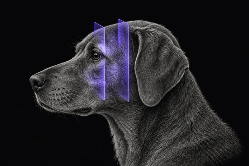
Why masticatory muscle myositis matters
Masticatory muscle myositis (MMM) is a focal inflammatory myopathy. In a review of 200 dogs with inflammatory myopathy diagnosed on muscle biopsy (Evans et al., Journal of Veterinary Internal Medicine, 2004), dogs were classified as having a generalised or a focal form, and masticatory muscle myositis and extraocular myositis made up the focal group. Common signs across that series included dysphagia, masticatory or generalised muscle atrophy and an inability to open the jaw.
The autoantibodies in MMM target type 2M fibres. As a 2022 case report of a young Cavalier King Charles Spaniel (Di Tosto et al., Frontiers in Veterinary Science) explains, the temporalis, masseter and pterygoid muscles contain these fibres; the digastricus does not. Swelling and pain are typical of the acute phase. In the chronic phase, atrophy dominates and muscle is replaced by fibrous tissue, and dogs treated at that stage may carry a worse prognosis for improvement.
When MRI is indicated
MRI is worth considering when a dog has trismus, jaw pain, swelling or atrophy of the temporal and masseter muscles, or eye signs that could come from the masticatory muscles.
-
Atrophy has more than one cause. In 63 dogs that had MRI for unilateral masticatory muscle atrophy (Milodowski et al., Journal of Veterinary Internal Medicine):
- a presumed trigeminal nerve sheath tumour was diagnosed in 30 (47.6%)
- other extra-axial masses in 13 (20.6%)
- no MRI abnormality was found in 18 (28.6%)
Most dogs with a presumed nerve sheath tumour, and all dogs with another extra-axial mass, had additional neurological deficits. The authors stress a neurological examination and MRI in these dogs.
-
Eye signs. In one dog with dynamic exophthalmos and lateral strabismus (Czerwinski et al., Veterinary Ophthalmology, 2015), CT showed a contrast-enhancing region of the masseter impinging on the orbit, more obvious with the mouth open. Serology and histology confirmed MMM.
Serology, imaging and biopsy
Serology is the usual first test. The 2M antibody test has been described as the diagnostic gold standard (Cauduro et al., Journal of the American Animal Hospital Association, 2013). A negative result needs care, though. In muscle from 53 dogs with MMM (Neumann and Bilzer, Muscle & Nerve, 2006), some dogs had no detectable intramuscular (26%) or circulating (36%) autoantibodies, yet still had CD8+ T-cell infiltrates and muscle fibre lesions. Where serology and imaging disagree, biopsy is worth discussing.
Masticatory muscle MRI protocol at a glance
The authors of the 2022 case report describe MRI as the modality best suited to early detection of affected muscles. Reported findings rely on contrast enhancement of the temporalis, masseter and pterygoid muscles, which also guides where to biopsy. When limb weakness or a high creatine kinase suggests generalised disease, including the neck and proximal limb muscles in the scan can be worth discussing.
What the radiologist looks at
| Modality | Findings reported in dogs with MMM |
|---|---|
| MRI | Widespread, symmetrical, inhomogeneously hyperintense areas in the masticatory muscles in two dogs. In a chronic juvenile case: severe symmetrical atrophy; temporalis, masseter and pterygoid muscles heterogeneously hyperintense on T2-weighted, FLAIR and STIR images, isointense on T1-weighted images, with strong diffuse contrast enhancement that spared the digastricus; regional lymph nodes enlarged. |
| CT | In 7 dogs (Reiter and Schwarz, Journal of the American Veterinary Medical Association, 2007): atrophy or swelling of all masticatory muscles except the digastricus (involved in 1 dog); pre-contrast hypoattenuation in 4 dogs; inhomogeneous contrast enhancement of the temporalis, masseter and pterygoid muscles in all dogs; enlarged head and neck lymph nodes in all but 1 dog. |
- Where to biopsy. In the CT series, muscle biopsies in 6 dogs were taken from the areas with the most obvious contrast enhancement, and all 6 had histological features of masticatory myositis. The same reasoning is usually applied to MRI: the report should name the muscle and region that enhance most, so the sample is taken from active disease rather than fibrotic tissue.
- Unilateral atrophy. The radiologist also looks for a nerve sheath tumour or another extra-axial mass, the most common MRI diagnoses in the 63-dog study.
What your report will include
Every Veheri case is read by a radiologist or neurologist, and when you order you can choose Diplomate-only reporting or the full specialist panel. Neurology reporting covers CT and MRI. You can chat directly with the specialist assigned to your case, and a simplified pet owner report helps you explain the findings to the owner.
When you take the report forward:
- Imaging shows inflamed muscle, not its cause. In the 200-dog review, inflammatory myopathy was associated with antibody titres against infectious diseases in 38 dogs, so infectious causes still need to be considered.
- Focal or generalised. Average creatine kinase (CK) and AST concentrations were significantly higher in the generalised forms than in the focal forms.
- Before the next anaesthetic. A Pug with MMM and trismus developed tongue protrusion under anaesthesia for diagnostic testing (Nanai et al., Veterinary Surgery, 2009). Venous congestion caused severe swelling, and a mandibular symphysiotomy was needed to return the tongue to the mouth. The authors advise monitoring tongue position and avoiding monitoring equipment on the tongue, such as a pulse oximeter probe, in these patients.
- Talking to the owner: dogs treated in the chronic, fibrotic phase may carry a worse prognosis for improvement.
What to include when you request an MRI report
- signalment: age, breed and sex
- duration of signs, and whether the muscles are swollen and painful or atrophied
- jaw range of motion, including trismus or an inability to close the mouth
- whether atrophy is unilateral or bilateral, and any other cranial nerve deficits
- eye signs such as exophthalmos or strabismus
- limb weakness and creatine kinase results
- 2M antibody and infectious disease results, and any treatment already given
How Veheri can help
Veheri works with veterinary radiologists and neurologists and reports MRI and CT studies remotely for clinics. For unilateral atrophy, see MRI of the trigeminal nerve in dogs, and for eye signs, see MRI of the orbit in dogs.
Sources
Each claim in this guide links to its source. The full citations:
- Evans J, Levesque D, Shelton GD. Canine inflammatory myopathies: a clinicopathologic review of 200 cases. J Vet Intern Med. 2004;18(5):679-91. doi:10.1892/0891-6640(2004)18<679:cimacr>2.0.co;2
- Di Tosto M, Callegari C, Matiasek K, et al. Case report: Atypical and chronic masticatory muscle myositis in a 5-month old Cavalier King Charles Spaniel. Clinical and diagnostic findings, treatment and successful outcome. Front Vet Sci. 2022;9:955758. doi:10.3389/fvets.2022.955758
- Milodowski EJ, Amengual-Batle P, Beltran E, et al. Clinical findings and outcome of dogs with unilateral masticatory muscle atrophy. J Vet Intern Med. 33(2):735-42 (published online 2018). doi:10.1111/jvim.15373
- Czerwinski SL, Plummer CE, Greenberg SM, et al. Dynamic exophthalmos and lateral strabismus in a dog caused by masticatory muscle myositis. Vet Ophthalmol. 2015;18(6):515-20. doi:10.1111/vop.12260
- Cauduro A, Paolo F, Asperio RM, et al. Use of MRI for the early diagnosis of masticatory muscle myositis. J Am Anim Hosp Assoc. 2013;49(5):347-52. doi:10.5326/JAAHA-MS-5915
- Reiter AM, Schwarz T. Computed tomographic appearance of masticatory myositis in dogs: 7 cases (1999-2006). J Am Vet Med Assoc. 2007;231(6):924-30. doi:10.2460/javma.231.6.924
- Neumann J, Bilzer T. Evidence for MHC I-restricted CD8+ T-cell-mediated immunopathology in canine masticatory muscle myositis and polymyositis. Muscle Nerve. 2006;33(2):215-24. doi:10.1002/mus.20456
- Nanai B, Phillips L, Christiansen J, et al. Life threatening complication associated with anesthesia in a dog with masticatory muscle myositis. Vet Surg. 2009;38(5):645-9. doi:10.1111/j.1532-950X.2009.00515.x
Related reading
More imaging guides
All imaging guides
MRI of the canine brachial plexus for peripheral nerve sheath tumours
How MRI finds peripheral nerve sheath tumours of the canine brachial plexus, what the radiologist looks for, and what imaging can say about grade and extent.
Brain MRI for dogs with seizures
Which dogs with seizures need brain MRI, what the evidence says about diagnostic yield, what the radiologist looks for and what your report will include.
MRI of the canine cervical spine for cervical spondylomyelopathy
How MRI characterises cervical spondylomyelopathy in dogs, what traction and flexion-extension studies add, and why compression alone does not prove the cause.