
Why the choice of imaging matters
Discospondylitis is an infection of the intervertebral disc and the adjacent vertebral endplates. In a multi-institutional study of 386 dogs (Van Hoof et al., Journal of Veterinary Internal Medicine, 2023):
- male dogs were overrepresented (236 of 386)
- L7–S1 was the most common site (97 of 362 dogs with site data)
- multifocal lesions were present in 134 of 362 dogs (37.0%)
Only 31 of 386 dogs (8.0%) had a temperature above 39.7 °C, so the authors caution that a normal temperature does not rule out infection.
Infection does not always start in the disc. In 33 dogs with Brucella canis discospondylitis (Long et al., Frontiers in Veterinary Science, 2022), MRI showed vertebral physitis or spondylitis without discitis in 7 of 18 dogs (39%).
Radiographs, CT or MRI
In 165 dogs that had at least one imaging modality at diagnosis, 55 (33.3%) had no radiographic evidence of discospondylitis but had lesions on CT, MRI or both. Four dogs had lesions on MRI alone. Agreement on whether disease was present was:
- fair between radiographs and CT (κ = 0.22)
- none to slight between radiographs and MRI (κ = 0.05)
- none to slight between CT and MRI (κ = 0.06)
Agreement on where the disease was located was much better, and substantial between CT and MRI (κ = 0.72).
| Modality | What the evidence shows |
|---|---|
| Radiographs | Can miss disease that CT or MRI detects. In B. canis cases, smooth, round, central endplate lysis (“hole punch” lesions) was seen on radiographs in 25 of 29 dogs (86%). |
| CT | In 41 dogs with MRI-confirmed disease, 63 discs (Gomes et al., Journal of Veterinary Internal Medicine, 2022): endplate involvement in 87.3%, mostly bilateral, with erosion and multifocal osteolysis; periosteal proliferation in 73%; vertebral body involvement in 66.7%. |
| MRI | In 13 dogs, 17 sites (Carrera et al., Veterinary Radiology & Ultrasound): MRI showed early lesions that radiography may not. |
Special groups
- Bulldogs. In English and French bulldogs (Grapes et al., Journal of Veterinary Internal Medicine, 2024), discospondylitis prevalence was 3.4 times higher in French bulldogs and 4.3 times higher in English bulldogs than in the hospital population. It was diagnosed next to a congenital vertebral body malformation in 80% of affected discs in French bulldogs and 50% in English bulldogs. Median age was about 1 year, against 7.3 years in other breeds.
- Fungal disease. In 11 dogs with mycotic discospondylitis (Okonji et al., Journal of Veterinary Internal Medicine, 2025), 7 were German Shepherd dogs and MRI showed several affected discs in 7. Fungal hyphae were found in urine sediment in 5 dogs and by CT-guided aspiration of the disc in 2.
Spinal MRI protocol at a glance
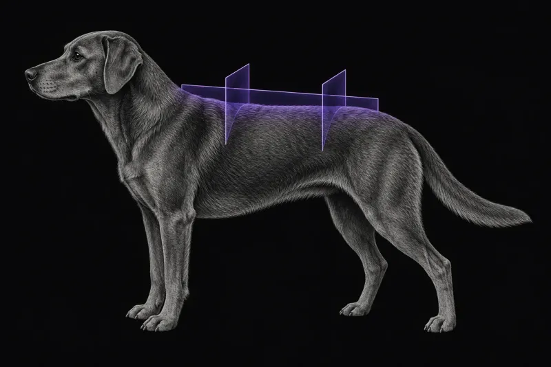
Several disc spaces may be affected, so a review of discospondylitis imaging (Ruoff et al., Veterinary Clinics of North America: Small Animal Practice) advises that imaging of the entire spine should be considered. On MRI, the 13-dog study described the lesions on T1-weighted, STIR and post-contrast images. When CT is used, all three planes must be reviewed, and equivocal cases may still need MRI.
What the radiologist looks at
- Disc and both endplates. In the 13-dog MRI study, two adjacent endplates and the disc were always involved. Endplates and adjacent marrow were T1-hypointense and STIR-hyperintense, with enhancement of endplates and paravertebral tissues in all dogs.
- Epidural extension. It was conspicuous after contrast at 15 sites (88.2%).
- Mimics. Endplate changes are not all infection. In 75 dogs with 76 endplate lesions on MRI (Gendron et al., Veterinary Radiology & Ultrasound, 2012), presumptive diagnoses were discospondylitis (29 dogs), fatty infiltration (26), reactive endplate changes (10), vertebral osteochondrosis (7) and intravertebral disc herniation (4). Irregular endplates, T2 or STIR hyperintensity, reduced T1 signal and contrast enhancement were seen in discospondylitis and in other non-fatty endplate changes. The authors advise careful evaluation of the adjacent structures when these features overlap.
- Every affected site. Multifocal disease is common, and a report that names the most accessible affected disc helps plan sampling.
What your report will include
Every Veheri case is read by a radiologist or neurologist, and when you order you can choose Diplomate-only reporting or the full specialist panel. Neurology reporting covers CT and MRI. You can chat directly with the specialist assigned to your case, and a simplified pet owner report helps you explain the findings to the owner.
When you take the report forward:
- Imaging does not name the organism. In the 386-dog study, blood cultures were positive in 38 of 143 dogs (27%), urine cultures in 77 of 277 (27.8%) and disc cultures in 26 of 63 (41%). The review recommends considering image-guided biopsy alongside blood and urine cultures and B. canis screening.
- Screen for Brucella whatever the reproductive status. Screening was positive in 9 of 136 dogs, including neutered dogs, and the authors suggest considering brucellosis regardless of reproductive status.
- Follow-up MRI needs care. In 25 dogs with a routine follow-up low-field MRI (de Freitas et al., Frontiers in Veterinary Science, 2022), 16 were asymptomatic, yet the two observers rated 20 (80%) and 18 (72%) of the dogs as having active disease. No follow-up MRI feature was associated with clinical status, so follow-up images are read together with the clinical picture.
What to include when you request a spinal MRI report
- signalment: age, breed and sex
- duration of pain, neurological deficits and temperature
- previous spinal surgery, trauma or corticosteroid therapy
- whether the dog is a bulldog or German Shepherd dog
- culture, urine and Brucella results available so far
- earlier radiographs or CT for comparison
How Veheri can help
Veheri provides veterinary teleradiology and teleneurology for X-ray, CT and MRI, with expert support for CT and MRI scanning protocols, positioning and acquisition. Related reading: MRI of the canine lumbosacral spine, CT of the canine thoracolumbar spine and a spinal CT protocol guide.
Sources
Each claim in this guide links to its source. The full citations:
- Van Hoof C, Davis NA, Carrera-Justiz S, et al. Clinical features, comparative imaging findings, treatment, and outcome in dogs with discospondylitis: A multi-institutional retrospective study. J Vet Intern Med. 2023;37(4):1438-1446. doi:10.1111/jvim.16785
- Long C, Burgers E, Copple C, et al. Brucella canis discospondylitis in 33 dogs. Front Vet Sci. 2022;9:1043610. doi:10.3389/fvets.2022.1043610
- Gomes SA, Targett M, Lowrie M. Computed tomography features of discospondylitis in dogs. J Vet Intern Med. 2022;36(6):2123-2131. doi:10.1111/jvim.16551
- Carrera I, Sullivan M, McConnell F, Gonçalves R. Magnetic resonance imaging features of discospondylitis in dogs. Vet Radiol Ultrasound. 52(2):125-31 (published online 2010). doi:10.1111/j.1740-8261.2010.01756.x
- Ruoff CM, Kerwin SC, Taylor AR. Diagnostic imaging of discospondylitis. Vet Clin North Am Small Anim Pract. 48(1):85-94 (published online 2017). doi:10.1016/j.cvsm.2017.08.007
- Gendron K, Doherr MG, Gavin P, Lang J. Magnetic resonance imaging characterization of vertebral endplate changes in the dog. Vet Radiol Ultrasound. 2012;53(1):50-6. doi:10.1111/j.1740-8261.2011.01861.x
- Grapes N, Bertram S, Gonçalves R, De Decker S. Prevalence of discospondylitis and association with congenital vertebral body malformations in English and French bulldogs. J Vet Intern Med. 2024;38(6):3138-3143. doi:10.1111/jvim.17209
- Okonji S, Di Paola A, Benini S, et al. Clinical signs, advanced diagnostic imaging findings, treatment, and outcome of mycotic discospondylitis in 11 dogs. J Vet Intern Med. 2025;39(3):e70097. doi:10.1111/jvim.70097
- de Freitas MI, Vettorato E, Scarpante E, et al. Retrospective preliminary assessment of routine follow-up low-field magnetic resonance imaging in dogs presumptively diagnosed with discospondylitis. Front Vet Sci. 2022;9:880038. doi:10.3389/fvets.2022.880038
Related reading
More imaging guides
All imaging guides
MRI of the canine brachial plexus for peripheral nerve sheath tumours
How MRI finds peripheral nerve sheath tumours of the canine brachial plexus, what the radiologist looks for, and what imaging can say about grade and extent.
Brain MRI for dogs with seizures
Which dogs with seizures need brain MRI, what the evidence says about diagnostic yield, what the radiologist looks for and what your report will include.
MRI of the canine cervical spine for cervical spondylomyelopathy
How MRI characterises cervical spondylomyelopathy in dogs, what traction and flexion-extension studies add, and why compression alone does not prove the cause.